
To aid in the diagnosis and confirmation of patients for whom a clinical suspicion of Strongyloides infection exists. Results for this test should not be used without correlation to clinical history or other data. Strongyloidiasis is caused by the intestinal nematode, Strongyloides, as it migrates from the skin to the intestines. These nematodes may exist as free-living larvae in warm, moist tropical climates, or as non-infective larvae that pass in the stool of infected individuals and become infective while in the soil of temperate environments. Strongyloides larvae enter the body by penetrating the skin and are carried through blood vessels to the lungs. The larvae travel from the lungs to the trachea and the pharynx, where they are swallowed and enter the intestines via the duodenum and upper jejunum.
S stercoralis has a complex lifecycle that begins with maturation to the infective filariform larva in warm, moist soil. The larvae subsequently penetrate exposed skin and migrate hematogenously to the lungs, from where they ascend the bronchial tree and are swallowed. Once in the small intestine, filariform larva matures into the adult worms that burrow into the mucosa. Gravid female worms produce eggs that develop into noninfectious rhabditiform larvae in the gastrointestinal tract and are eventually released in the stool. The time from dermal penetration to appearance of Strongyloides in stool samples is approximately 3 to 4 weeks.
The most common manifestations of infection are mild and may include epigastric pain, mild diarrhea, nausea, and vomiting. At the site of filariform penetration, skin may be inflamed and itchy-this is referred to as "ground itch." Migration of the larva through the lungs and up the trachea can produce a dry cough, wheezing, and mild hemoptysis. Eosinophilia, though common among patients with strongyloidiasis, is not a universal finding, and the absence of eosinophilia cannot be used to rule-out infection.
In some patients, particularly those with a depressed immune system, the rhabditiform larvae may mature into the infectious filariform larvae in the gastrointestinal tract and lead to autoinfection. The filariform larvae subsequently penetrate the gastrointestinal mucosa, migrate to the lungs, and can complete their lifecycle. Low-level autoinfection can maintain the nematode in the host for years to decades. Among patients who become severely immunocompromised, however, autoinfection may lead to hyperinfection and fatal disseminated disease. Hyperinfection has also been associated with underlying human T-cell lymphotropic virus type 1 infection. Uncontrolled, the larvae can disseminate to the lungs, heart, liver, and central nervous system. Septicemia and meningitis are common in cases of Strongyloides hyperinfection due to seeding of the bloodstream and central nervous system with bacteria originating from the gastrointestinal tract.
Strongyloidiasis is caused by Strongyloides stercoralis, a nematode endemic to tropical and subtropical regions worldwide. S stercoralis is also prominent in the southeastern United States, including in rural areas of Kentucky, Tennessee, Virginia, and North Carolina. A small series of epidemiological studies in the United States identified that 0% to 6.1% of individuals sampled had antibodies to S stercoralis.
References:
Ramanathan R, Burbelo PD, Groot S, et al: A luciferase immunoprecipitation systems assay enhances the sensitivity and specificity of diagnosis of Strongyloides stercoralis infection. J Infect Dis. 2008;198(3):444-451
Starr MC, Montgomery SP: Soil-transmitted Helminthiasis in the United States: a systematic review-1940-2010. Am J Trop Med Hyg. 2011;85(4):680-684
Krolewiecki AJ, Ramanathan R, Fink V, et al: Improved diagnosis of Strongyloides stercoralis using recombinant antigen-based serologies in a community-wide study in northern Argentina. Clin Vaccine Immunol. 2010;17(10):1624-1630
Centers for Disease Control and Prevention. Global Health. Division of Parasitic Diseases and Malaria: Parasites-Strongyloides: Epidemiology and Risk Factors. CDC; www.cdc.gov/parasites/strongyloides/epi.html
 Strongyloides IgG Antibody
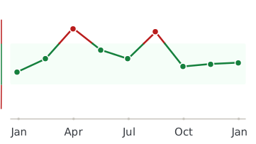
Every lab result you’ve ever had, charted and explained.
Healthmatters is a tracker for your lab results. Upload a report from any lab, any country — every marker charted and explained.
Start with my report →
All our content is backed by peer-reviewed studies, academic research, and trusted medical sources. We’re committed to accuracy and transparency — see our editorial policy for details.