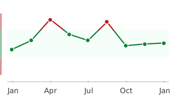
Magnesium (Mg) is an essential element with both electrolyte and enzyme-activator functions. However, neither of these functions takes place in hair. Body excess of Magnesium is rare but may occur from excessive oral or parenteral supplementation or as a result of renal damage or insufficiency.
If one rules out external contamination of hair as a result of recent hair treatment, elevated hair Magnesium is more likely to indicate maldistribution of the element. Physiological Magnesium dysfunction may or may not be present. Maldistribution of Magnesium can occur as a result of chronic emotional or physical stress, toxic metal or chemical exposure, physiological imbalance of calcium and phosphorus, bone mineral depletion, and renal insufficiency with poor clearance of Magnesium (and other metabolites). Elevated hair Magnesium has been correlated with hypoglycemia and an inappropriately low ratio of dietary Ca : P.
Magnesium status can be difficult to assess; whole blood and packed blood red cell Magnesium levels are more indicative than serum/plasma levels.
Amino acid analysis can be helpful in showing rate-limited steps that are Magnesium-dependent (e.g. phosphorylations).
Low hair Magnesium (Mg) levels may be indicative of Mg deficiency, but this has not been unequivocally demonstrated. When hair Mg is low, dietary intake and malabsorption should be considered. Mg is an essential element/electrolyte that is necessary for the activity of many important enzymes. Low hair Mg may or may not be associated with physiological dysfunction.
Causes of Mg deficiency include: consumption of a ”junk food” diet or Mg-deficient foods, intestinal malabsorption, hypocalcemia with decreased Mg retention, chemical toxicity with renal wasting, alcoholism, alkalosis, prolonged diarrhea/laxative abuse, and iatrogenic causes (digoxin therapy, occasionally from oral contraceptives, hypercalcemic drugs, gentamicin, neomycin).
Symptoms of Mg deficiency include: muscle twitching, cramps, tremor or muscle spasms, paresthesia, and mental depression. Low Mg status is associated with arrythmias and increased cardiovascular risk.
Mg status can be difficult to assess; whole blood and packed red cell levels are more indicative than serum/plasma levels. Amino acid analysis can be helpful in showing rate-limited steps that are Mg-dependent such as phosphorylations. Taurine deficiency is often associated with urinary loss of Mg. The Mg challenge method may be indicative: baseline 24-hour urine Mg measurement, followed by 0.2 mEq/Kg intravenous mg, followed by 24-hour Mg measurement. A deficiency is judged to be present if less than 80% of the administrered Mg is excreted in the urine.

 Magnesium