
QUICK ANSWER
MCH (Mean Corpuscular Hemoglobin) measures the average amount of hemoglobin in each red blood cell. More hemoglobin means more oxygen-carrying capacity per cell.
Normal range: 27–33 pg (picograms per cell); some labs use 26.6–33.0 pg
| MCH result | What it generally means |
|---|---|
| Below 26 pg | Low — iron deficiency or thalassemia; evaluate with ferritin and MCV |
| 26–27 pg | Borderline low — mild iron deficiency or early depletion possible |
| 27–33 pg | Normal |
| 33–35 pg | Mildly high — often B12/folate deficiency, alcohol, or lab variation |
| Above 35 pg | Elevated — evaluate for macrocytic anemia; check B12, folate, thyroid, liver |
Key principle: MCH and MCV almost always move together. High MCH = large red blood cells. Low MCH = small red blood cells. If MCV and MCH disagree significantly, repeat testing or further evaluation may be needed.
WHAT DOES MCH STAND FOR?
MCH stands for Mean Corpuscular Hemoglobin — the average mass of hemoglobin in each individual red blood cell:
- Mean — average across the red blood cells in the sample
- Corpuscular — relating to red blood cells (corpuscle = small body)
- Hemoglobin — the oxygen-carrying protein inside each cell
MCH is reported in picograms (pg) — trillionths of a gram — and measures total hemoglobin content per cell, not concentration. MCHC measures concentration; MCH measures total amount.
YOUR SPECIFIC MCH NUMBER — WHAT DOES IT MEAN?
The most commonly searched MCH values are in the low-normal to mildly high range:
| MCH value (pg) | Typical interpretation |
|---|---|
| Below 25 | Significantly low — evaluate for iron deficiency anemia or thalassemia |
| 25–26 | Low — iron deficiency likely; check ferritin, MCV, RDW |
| 26–26.9 | Borderline to mildly low; evaluate in context of MCV and ferritin |
| 26.6 | Lower limit of some lab reference ranges — borderline; check other CBC indices |
| 27–33 | Normal range |
| 33–33.9 | Mildly elevated — at or just above upper limit; often B12/folate or alcohol |
| 33.2 | Mildly above reference range; frequently B12, folate, alcohol, or liver |
| 34–35 | Moderately elevated — investigate for macrocytic cause |
| Above 35 | Significantly elevated — macrocytic anemia likely; clinical evaluation warranted |
Is MCH 33 high? 33 pg is at or just above the upper limit of most reference ranges (27–33 pg). It is mildly elevated — often benign in isolation, but warrants checking B12, folate, and MCV.
Is MCH 26.6 low? It depends on the lab. 26.6 pg is within the normal range for labs using a lower limit of 26.6 pg. For labs using 27 pg as the lower limit, it is mildly low. Check the reference range on your own lab report.
WHAT DOES HIGH MCH MEAN?
High MCH (above ~33 pg) means each red blood cell contains more hemoglobin than average. This almost always occurs when red blood cells are larger than normal — a condition called macrocytosis. High MCH and high MCV nearly always appear together.
Common causes of high MCH:
| Cause | Notes |
|---|---|
| Vitamin B12 deficiency | Most common cause of macrocytic anemia; MCH and MCV both elevated |
| Folate (folic acid) deficiency | Second most common macrocytic cause — often associated with poor diet or alcohol use |
| Alcohol use | Alcohol is directly toxic to red blood cell production; MCH elevation is a sensitive alcohol marker |
| Liver disease | Any cause of liver disease can raise MCH |
| Hypothyroidism | Underactive thyroid slows red blood cell maturation |
| Certain medications | Methotrexate, hydroxyurea, and some antiretrovirals can cause macrocytosis |
| Reticulocytosis | Large young red blood cells (reticulocytes) released during recovery from blood loss or hemolysis |
| Normal variation | Mild elevation (33–34 pg) with otherwise normal CBC is sometimes normal variation |
High MCH but everything else normal: A mildly elevated MCH (33–34 pg) with normal hemoglobin, normal MCV, and no symptoms is often benign. It most commonly reflects mild B12 or folate suboptimality, recent or ongoing alcohol intake, or normal biological variation. If MCV is also elevated, investigation for macrocytic cause is more warranted. If MCH is mildly high but MCV is normal, repeat testing and review of alcohol intake is usually the first step.
WHAT DOES LOW MCH MEAN?
Low MCH (below ~27 pg) means each red blood cell contains less hemoglobin than normal. This almost always occurs when red blood cells are smaller than normal — microcytosis. Low MCH and low MCV nearly always appear together.
Common causes of low MCH:
| Cause | Notes |
|---|---|
| Iron deficiency | Most common cause worldwide — insufficient iron reduces hemoglobin production |
| Iron deficiency anemia | Established iron deficiency with low hemoglobin; MCH, MCV, and hemoglobin all typically low |
| Thalassemia trait | Inherited structural hemoglobin abnormality; MCH and MCV both low, but RBC count is normal or high |
| Chronic blood loss | Heavy menstrual bleeding, GI bleeding — progressive iron depletion lowering MCH |
| Anemia of chronic disease | Chronic inflammation reduces iron availability; MCH may fall |
Low MCH with normal hemoglobin: A low MCH with normal hemoglobin often represents early iron depletion before anemia develops. Ferritin is the most sensitive test to confirm or exclude iron deficiency at this stage. Thalassemia trait also characteristically produces low MCH with preserved hemoglobin.
MCH AND MCV — COMBINED PATTERN INTERPRETATION
MCH and MCV are the most closely linked CBC indices — they rise and fall together in the great majority of cases. Interpreting them together identifies the anemia type:
| MCH | MCV | RDW | Most likely pattern |
|---|---|---|---|
| Low | Low | High | Iron deficiency anemia — most common |
| Low | Low | Normal | Thalassemia trait — RBC count usually normal or high |
| Normal | Normal | Normal | Normal CBC |
| High | High | Normal or elevated | Macrocytic anemia — check B12, folate, alcohol, liver, thyroid |
| High | High | Very high | Mixed deficiency or hemolytic process |
| High | High | High (with clinical context of recent blood loss or hemolysis) | Reticulocytosis — large young red blood cells released during recovery from blood loss or hemolysis; MCH and MCV transiently elevated |
| High | Normal | Normal | Mild MCH elevation — often variation, alcohol, or early macrocytosis |
| Low | Normal | Normal | Uncommon — early iron deficiency before MCV falls; check ferritin |
The thalassemia pattern: Low MCH + low MCV + normal RBC count (or high RBC) + normal RDW + normal ferritin = thalassemia trait strongly favored. This is in contrast to iron deficiency, where RBC count falls alongside MCH and MCV, and ferritin is low. Low MCH + low MCV + normal/high RBC count + normal ferritin is one of the most classic laboratory patterns of thalassemia trait and is particularly useful for distinguishing it from iron deficiency when the two conditions look similar on initial CBC.
MCH VS MCV VS MCHC — WHAT IS THE DIFFERENCE?
| Marker | What it measures | Units | Normal range |
|---|---|---|---|
| MCH | Average total amount of hemoglobin per red blood cell | pg (picograms) | 27–33 pg |
| MCV | Average size (volume) of red blood cells | fL (femtoliters) | 79–97 fL |
| MCHC | Average concentration of hemoglobin inside each red blood cell | g/dL | 32–36 g/dL |
MCH vs MCHC — the key difference: MCH measures how much hemoglobin is in a cell regardless of cell size. MCHC measures how densely packed that hemoglobin is. In a large red blood cell (high MCV), MCH may be high while MCHC is normal — the cell is big and carries more hemoglobin, but it's not necessarily more concentrated. MCHC is considered more specific for true hypochromia.
Why MCH and MCV move together: In iron deficiency, small cells are produced with less hemoglobin — both MCV and MCH fall. In B12/folate deficiency, large cells are produced with more hemoglobin — both rise. MCHC often remains normal in macrocytosis.
FERRITIN + MCH INTERPRETATION
| MCH | Ferritin | Most likely explanation |
|---|---|---|
| Low | Low | Iron deficiency — depleted stores directly reducing hemoglobin per cell |
| Low | Normal | Thalassemia trait, or early iron deficiency with preserved ferritin |
| Low | High | Anemia of chronic disease — iron sequestered by inflammation |
| Normal | Low | Early iron deficiency before MCH has fallen — check MCV and RDW |
| Normal | Normal | Iron deficiency unlikely — evaluate other causes if symptoms present |
| High | Normal | B12/folate deficiency, alcohol use, or liver disease more likely — ferritin is not typically low in macrocytic anemia |
Ferritin below 30 ng/mL is strongly supportive of iron deficiency. A normal MCH does not rule out iron deficiency — ferritin depletes before MCH changes.
MOST COMMON MCH RESULTS
| MCH result (pg) | Typical interpretation |
|---|---|
| 26.6 | Borderline low — at the lower limit of some lab reference ranges; mildly below others (lower limit 27.0) |
| 26 | Mildly low — check ferritin and MCV; likely early iron depletion if MCV is also low |
| 27 | Lower end of normal |
| 33 | Upper end of normal or mildly elevated — check MCV; evaluate for B12/folate if persistent |
| 33.2 | Mildly elevated — most commonly benign; review alcohol, B12, folate |
| 35 or above | Macrocytic evaluation warranted — check B12, folate, liver function, thyroid, and medications |
IS HIGH MCH DANGEROUS?
MCH does not have an emergency threshold the way creatine kinase (CK) or potassium does. Clinical significance depends on degree, trend, MCV, hemoglobin, and symptoms:
| MCH pattern | Clinical significance |
|---|---|
| 33–35 pg, everything else normal | Usually mild — monitor; check B12 and folate if persistent |
| 35–37 pg | Evaluate for B12 deficiency, folate deficiency, alcohol, liver disease, hypothyroidism |
| Above 37 pg | Significant macrocytosis — clinical evaluation warranted; check full panel including B12, folate, LDH, smear |
| High MCH + low hemoglobin (anemia) | Clinical evaluation needed — macrocytic anemia requires diagnosis and treatment |
| High MCH + neurological symptoms (tingling, memory changes) | Prompt B12 evaluation — B12 deficiency can cause neurological damage even before anemia develops |
A mildly elevated MCH in isolation rarely requires urgent action. The most common serious underlying cause is B12 deficiency, which is treatable — and neurological damage can be prevented if caught early.
DOES MCH CHANGE WITH AGE?
MCH reference ranges are relatively stable across adult demographic groups, but some age-related variation exists:
| Group | Typical pattern |
|---|---|
| Children | Pediatric reference ranges differ significantly — lower than adult ranges in infants, increasing with age |
| Adult women | Standard adult range (27–33 pg) |
| Adult men | Standard adult range (27–33 pg) — sex differences in MCH are minimal |
| Pregnancy | Mild shifts possible — folate demands increase during pregnancy, which can affect MCH |
| Older adults (65+) | Mild increases more common due to higher rates of B12 deficiency and chronic disease in this group |
The most clinically important demographic pattern is in older adults: B12 deficiency becomes more common with age (due to reduced gastric acid and intrinsic factor), and a mildly elevated MCH in an older adult warrants B12 testing even if the elevation is small.
WHY TRENDS MATTER MORE THAN ONE RESULT
MCH is most informative as a trend over time. Users with longitudinal CBC data on HealthMatters can apply this framework:
| Pattern | Clinical meaning |
|---|---|
| Stable MCH of 33 pg over several years | Often individual baseline variation — lower clinical concern |
| MCH declining: 30 → 26 pg over multiple tests | Progressive iron depletion — evaluate ferritin and address cause |
| MCH rising: 28 → 33 pg over time | Macrocytosis developing — check B12, folate, alcohol, thyroid |
| MCH improving after B12 or folate supplementation | Supports deficiency as the cause — treatment response confirms diagnosis |
| MCH persistently low despite adequate iron therapy | Consider thalassemia trait — thalassemia does not respond to iron and MCH remains low |
A single mildly abnormal MCH on one test is very different from a trend. Falling MCH across 3–4 CBCs is more significant than a single mildly low result — even if the endpoint looks similar.
WHEN SHOULD YOU FOLLOW UP?
Most MCH abnormalities do not require urgent action. Consider discussing your result with a healthcare professional if:
| Situation | Suggested action |
|---|---|
| MCH below 27 pg on repeat testing | Evaluate with ferritin, MCV, and RDW — rule out iron deficiency or thalassemia |
| MCH above 35 pg | Check B12, folate, liver function, thyroid, alcohol history, and medications |
| MCH changing significantly over time | Any meaningful trend (up or down) warrants investigation regardless of current absolute value |
| Anemia present (low hemoglobin) alongside abnormal MCH | Clinical evaluation needed — type of anemia should be determined and treated |
| High MCH + neurological symptoms (tingling, numbness, memory changes) | Prompt B12 evaluation — neurological damage can occur before anemia develops |
| Ferritin, B12, or folate already known to be abnormal | Treat the underlying deficiency and monitor MCH for response |
| MCH persistently low despite iron supplementation | Consider thalassemia trait — hemoglobin electrophoresis may be appropriate |
A single mildly abnormal MCH result without symptoms or other CBC abnormalities usually requires no immediate action — monitoring on the next routine CBC is often sufficient.
 Mean Corpuscular Hemoglobin (MCH)
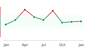
Every lab result you’ve ever had, charted and explained.
Healthmatters is a tracker for your lab results. Upload a report from any lab, any country — every marker charted and explained.
Start with my report →
Frequently Asked Questions
Can MCH be normal if iron is low?
What does high MCH mean in a blood test?
What does low MCH mean in a blood test?
What does MCH stand for in a blood test?
What does MCH 33 mean?
What does MCH 26.6 mean?
Can MCH be high if everything else is normal?
What is the difference between MCH and MCHC?
Can iron deficiency cause high MCH?
All our content is backed by peer-reviewed studies, academic research, and trusted medical sources. We’re committed to accuracy and transparency — see our editorial policy for details.