
Appear CSF

Sample type: Cerebrospinal fluid (CSF)

Other names: Appearance, CSF

Clear
A clear appearance of cerebrospinal fluid (CSF) generally indicates a normal or non-pathological state, as CSF is typically clear and colorless. This clarity suggests the absence of red blood cells (which could indicate bleeding or hemorrhage), high protein content (often associated with inflammation or infection), or other cellular debris that can cloud the fluid. A clear CSF is often seen in cases where there is no infection, bleeding, or significant inflammatory response within the central nervous system. However, it's important to note that a clear appearance does not entirely rule out all neurological conditions. For a comprehensive diagnosis, the clarity of CSF should be considered alongside other CSF parameters like cell count, glucose and protein levels, and specific diagnostic tests, such as those for infections or malignancies.
Yellow
If the cerebrospinal fluid (CSF) appearance is yellow, it typically indicates the presence of xanthochromia. Xanthochromia is the yellow discoloration of CSF and is often associated with specific medical conditions. Subarachnoid Hemorrhage (SAH): Yellow CSF can be a significant indicator of subarachnoid hemorrhage (SAH), a type of bleeding that occurs in the space surrounding the brain. It is one of the key clinical features of SAH. Xanthochromia and High Protein Levels: Xanthochromia can also occur in cases of high protein levels in the CSF. This can be due to various medical conditions and may require further evaluation to determine the underlying cause. Red Flag: The presence of yellow CSF, or xanthochromia, is considered a red flag in clinical practice, especially in the context of SAH. It prompts healthcare professionals to investigate the patient's condition further to determine the cause of the discoloration. It's important to note that while xanthochromia is often associated with SAH, it is not pathognomonic, meaning it can occur in other situations as well.
Orange
If the appearance of cerebrospinal fluid (CSF) is orange, it may indicate a medical condition called xanthochromia. Xanthochromia refers to the yellow or orange discoloration of CSF and is typically due to the presence of certain substances in the fluid. This discoloration can result from the breakdown of red blood cells or the presence of substances such as oxyhemoglobin, methemoglobin, or bilirubin in the CSF. Xanthochromia is often associated with various medical conditions, including subarachnoid hemorrhage, where there is bleeding into the space surrounding the brain and spinal cord. It can also be a sign of other neurological disorders.
Pink
If the cerebrospinal fluid (CSF) appears pink, it typically indicates the presence of xanthochromia, which is a discoloration of the CSF due to the presence of bilirubin. Xanthochromia can result from various medical conditions, most commonly subarachnoid hemorrhage (bleeding into the space surrounding the brain). It is a valuable diagnostic sign that helps healthcare professionals assess the presence of blood in the CSF. A pink CSF appearance can be observed when the CSF is examined after a traumatic lumbar puncture (a "traumatic tap"), as blood from the needle insertion may mix with the CSF. To differentiate between xanthochromia caused by a medical condition and that resulting from a traumatic tap, healthcare providers may use specific criteria and laboratory tests.

Cerebrospinal fluid (CSF) is a clear, watery liquid that flows around the brain and spinal cord, surrounding and protecting them. CSF testing is performed to evaluate the level or concentration of different substances and cells in CSF in order to diagnose conditions affecting the brain and spinal cord (central nervous system).

The appearance of the sample of CSF is usually compared to a sample of water.

Color of the fluid - normal is clear and colorless. Changes in the color of the CSF are not diagnostic but may point to additional substances in the fluid. Yellow, orange, or pink CSF may indicate the breakdown of blood cells due to bleeding into the CSF or the presence of bilirubin. Green CSF may also sometimes be seen with bilirubin or infection.

Turbidity - cloudy or turbid CSF may indicate the presence of white or red blood cells, microbes, or an increase in protein levels.

Viscosity - normal CSF will have the same consistency as water. CSF that is “thicker” may be seen in people with certain types of cancers or meningitis.

Conditions associated with changes in the appearance of CSF include the following:

Infectious meningitis – Turbid, milky, cloudy CSF samples

Hemorrhage or traumatic tap – Xanthochromic CSF samples with increased hemoglobin

Kernicterus - Xanthochromic CSF samples with increased bilirubin

Meningeal melanosarcoma – Xanthochromic CSF samples with increased melanin

Disorders affecting blood-brain barrier - Cloudy CSF samples with increased proteins (above 150 mg/dL) and albumin and IgG

Xanthochromia is the term used for any kind of discoloration of CSF (pink, yellow, orange).

Multiple conditions are associated with xanthochromia:

traumatic tap, presence of carotene, melanoma and increased bilirubin concentration (bilirubin concentration will also be elevated in serum and patients are often jaundiced) due to liver diseases, hemolytic diseases (also increased free hemoglobin concentration) and inborn errors of metabolism.

Note that normal CSF samples should be colorless, clear, like water. If CSF samples are centrifuged immediately, xanthochromia due to traumatic tap should not occur. However, if CSF samples are carefully centrifuged immediately and the supernatant is still xanthochromic, this indicates that bleeding may have occurred 2-4 hours before sample collection. Furthermore, in about 10% of patients with subarachnoid hemorrhage, the CSF samples might be clear if the samples are collected 12 hours after the hemorrhage occurred.

Typically, high levels of oxyhemoglobin occur in CSF fluid obtained through a traumatic lumbar puncture, in which red blood cells enter the subarachnoid space via direct needle puncture. This frustrates the ability to determine whether xanthochromia, and thus subarachnoid hemorrhage, is present.

Appear CSF
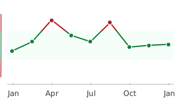
Healthmatters dashboard showing biomarker trend graph

Every lab result you’ve ever had, charted and explained.

Healthmatters is a tracker for your lab results. Upload a report from any lab, any country — every marker charted and explained.

Start with my report →
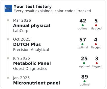
Beyond Appear CSF — 10,000+ markers explained
One Appear CSF result tells you where you are. Your history tells you where you’re heading. Upload your past lab reports and see this marker — and every marker on them —every marker charted against your own lab’s ranges.
Start with this report →
Healthmatters timeline preview showing biomarker trends over time
Clinical Sources & References

All our content is backed by peer-reviewed studies, academic research, and trusted medical sources. We’re committed to accuracy and transparency — see our editorial policy for details.

Article Review Log
Written by Healthmatters Editorial Team
Healthmatters Lifetime Plan

Pay $250. Track your health forever

One payment. No renewals. Yours for life.

10,000+ biomarkers explained · trends over time

See other plans →
Secure checkout via Stripe · GDPR & HIPAA
We accept results from any lab
LabCorp Quest Diagnostics Genova Diagnostics DUTCH Test Doctor's Data Vibrant America ZRT Laboratory Cyrex Labs + any other lab

What members are saying

★★★★★ 10,000+ happy members
★★★★★

"Watching my numbers move over the years — instead of forgetting last time's results — completely changed how I manage my health."

Daniel
Member since 2022
★★★★★

"Living across several countries made keeping my lab results organized a real challenge. Healthmatters brought my results from the USA, Spain, and Germany together — 20 years of history, with long-term trends instead of scattered reports."

Ben
Member since 2021 · tracks across countries
★★★★★

"Standard lab ranges are too wide for how I practice — 'normal' isn't optimal. I set my own bands, so a client sees yellow before a marker goes officially out of range. That's the conversation I want to be having."

Dr. Alexandra
Functional medicine · 6 yrs on Pro