Transferrin Blood Test: What It Measures, Normal Range, and What Low or High Levels Mean
Other names: Transferrin, Serum Transferrin, S-Transferrin, S Transferrin, Transferrin Serum, Plasma Transferrin, TRFN, TRF Blood Test, T'Ferrin, T Ferrin, Transferrin [Mass/Volume] in Serum or Plasma, Transferrin Protein, Siderophilin, Transferrin g/L, Transferrin mg/dL, Blood Transferrin, Transferrin Lab Test, Transferrin Blood Work
WHAT IS A TRANSFERRIN BLOOD TEST?
If your lab report shows "Transferrin," "S-Transferrin," "TRFN," or "T'Ferrin":
- This measures how much of the iron-transport protein transferrin is in your blood
- It is produced by the liver and reflects both iron status and liver function
- Low transferrin most commonly means iron overload, liver disease, chronic inflammation, or malnutrition
- High transferrin most commonly means iron deficiency — the liver makes more to capture scarce iron
- The result appears in mg/dL (US labs) or g/L (most international labs); 200 mg/dL = 2.0 g/L
Quick interpretation:
| Result | Usually means |
|---|---|
| Low (< 200 mg/dL / < 2.0 g/L) | Reduced production (liver disease, malnutrition, inflammation) or iron overload saturating binding sites |
| Normal (200–390 mg/dL / 2.0–3.9 g/L) | Adequate iron transport capacity; normal liver protein production |
| High (> 390 mg/dL / > 3.9 g/L) | Iron deficiency — liver produces more transferrin when iron stores are low |
If you only remember four things about transferrin:
- High transferrin usually suggests iron deficiency — the liver produces more to capture scarce iron
- Low transferrin usually reflects liver disease, inflammation, malnutrition, or iron overload — not iron deficiency
- Never interpret transferrin without ferritin and transferrin saturation — the pattern across all three is what identifies the diagnosis
- Ferritin almost always changes before hemoglobin — by the time hemoglobin falls, iron deficiency is advanced
LABEL DECODER — WHAT YOUR REPORT MIGHT SHOW
| Label on report | What it means |
|---|---|
| Transferrin | Standard label — serum or plasma transferrin concentration |
| S-Transferrin | "S" = serum; same measurement |
| S Transferrin | Variant formatting of S-Transferrin |
| Serum Transferrin | Explicit specimen designation |
| T'Ferrin | Abbreviated; used by some UK and Australian labs |
| T Ferrin | Alternative abbreviation |
| TRFN | Common lab shortcode |
| TRF | Shorter lab shortcode |
| Transferrin, Serum | Formal test name with specimen |
| Transferrin [Mass/volume] in Serum or Plasma | Full LOINC-style label |
| Transferrin Protein | Less common; same measurement |
| Siderophilin | Historical name for transferrin; occasionally appears on older reports |
| Transferrin g/L | International unit format |
| Transferrin mg/dL | US unit format |
All of these measure the same thing: the concentration of the iron-transport protein transferrin in blood.
UNIT CONVERSION — mg/dL AND g/L
US laboratories typically report transferrin in mg/dL. Most international labs (UK, Australia, Canada, EU) use g/L.
Conversion: mg/dL ÷ 100 = g/L. g/L × 100 = mg/dL.
| mg/dL | g/L | Interpretation |
|---|---|---|
| < 150 | < 1.5 | Severely low; marked protein deficiency, severe liver disease, or iron overload |
| 150–199 | 1.5–1.99 | Low; below reference range at most labs |
| 200 | 2.0 | Lower boundary of normal |
| 200–390 | 2.0–3.9 | Normal range |
| 250–300 | 2.5–3.0 | Mid-normal; well within reference range |
| 350–390 | 3.5–3.9 | Upper-normal; borderline elevated |
| 391–450 | 3.91–4.5 | Mildly elevated; evaluate for iron deficiency |
| > 450 | > 4.5 | Clearly elevated; iron deficiency likely; check serum iron and ferritin |
NORMAL RANGES
Many laboratories use a reference interval of approximately 200–390 mg/dL (2.0–3.9 g/L), though ranges vary slightly by laboratory and method.
| Population | Normal range (mg/dL) | Normal range (g/L) |
|---|---|---|
| Adults (general) | 200–390 mg/dL | 2.0–3.9 g/L |
| Pregnancy | Higher; up to 450–500 mg/dL | 4.5–5.0 g/L |
| Children | Similar to adults; varies by age | |
| Elderly | May trend slightly lower due to reduced liver production |
Note on g/L values: International labs often express results to one decimal place in g/L. Common results include 1.8, 1.9, 2.0, 2.1, 2.2, 2.3, 2.4, 2.5, 2.6, 2.7, 2.8, 2.9, 3.0, 3.1, 3.2, 3.5, 3.7 g/L. See the individual value lookup section below.
"MY TRANSFERRIN IS X" — INDIVIDUAL VALUE LOOKUP
Results in g/L can be converted to mg/dL by multiplying by 100 (e.g., 2.1 g/L = 210 mg/dL).
| Transferrin | What it means |
|---|---|
| < 1.5 g/L (< 150 mg/dL) | Severely low. Consistent with severe liver disease, critical illness, or marked iron overload. This result requires clinical evaluation — it does not diagnose iron deficiency. |
| 1.5–1.7 g/L (150–170 mg/dL) | Clearly low. Below the reference range at most labs. Check ferritin and CRP first: high ferritin + low transferrin = likely iron overload; high CRP + low transferrin = likely inflammation suppressing production. |
| My transferrin is 1.8 g/L | Slightly below normal. By itself this does not diagnose iron overload or liver disease. The next question is whether ferritin is high (iron overload) or CRP is elevated (inflammation). A repeat test after resolving acute illness is often appropriate. |
| My transferrin is 1.9 g/L | Borderline low — just below the typical lower limit of 2.0 g/L. Interpret alongside iron, ferritin, CRP, and albumin. If all others are normal, this may simply be a low-normal variation. |
| My transferrin is 2.0 g/L | At the lower boundary of normal at most laboratories. This result does not indicate iron deficiency; it is more consistent with adequate iron stores or mildly reduced liver protein production. |
| My transferrin is 2.1 g/L | Normal. If ferritin, serum iron, and transferrin saturation are also normal, this result is entirely reassuring and requires no action. |
| 2.2–2.4 g/L (220–240 mg/dL) | Normal. |
| My transferrin is 2.5 g/L | Normal mid-range. Reassuring if the remainder of the iron panel is also normal. |
| 2.6–2.9 g/L (260–290 mg/dL) | Normal. |
| My transferrin is 3.0 g/L | Normal mid-to-upper range. |
| 3.1–3.4 g/L (310–340 mg/dL) | Normal. |
| My transferrin is 3.5 g/L | Normal upper range. If ferritin is low or borderline, this could represent early iron deficiency — the liver starts making more transferrin before anemia develops. |
| My transferrin is 3.7 g/L | High-normal to borderline elevated. If ferritin is low, this often represents early iron deficiency before anemia develops. If ferritin is normal and iron is normal, this may be a normal variation or pregnancy-related. |
| My transferrin is 3.9 g/L | At the upper boundary of normal at most labs. Check serum iron and ferritin if not already tested. |
| 4.0–4.5 g/L (400–450 mg/dL) | Mildly elevated. The liver is producing more transferrin, most likely because iron stores are low. Check ferritin and transferrin saturation. |
| > 4.5 g/L (> 450 mg/dL) | Clearly elevated. Iron deficiency is the most common cause. Pregnancy and OCP use are physiological causes. Check serum iron, ferritin, and transferrin saturation to confirm the mechanism. |
When should I be concerned about my transferrin result?
| Result | Urgency | What to do |
|---|---|---|
| < 1.5 g/L | Prompt workup needed | Evaluate for liver failure, critical illness, or severe iron overload; not a result to wait on |
| 1.5–1.7 g/L | See a physician | Below normal range; evaluation of liver function, nutrition, and iron status warranted |
| 1.8–1.9 g/L | Review with doctor, usually not urgent | Mildly below normal; interpret with iron panel and CRP; repeat after acute illness if applicable |
| 2.0–3.9 g/L | Normal; no action needed unless iron panel is abnormal | |
| 4.0–4.2 g/L | Likely iron deficiency; evaluate soon | Check serum iron and ferritin; not an emergency but warrants follow-up |
| 4.5–5.0 g/L | Evaluate soon | Iron deficiency or pregnancy; ferritin and iron saturation needed |
| > 5.0 g/L | Evaluate | Significant elevation; iron deficiency workup including source investigation |
WHAT DOES LOW TRANSFERRIN MEAN?
Low transferrin (below 200 mg/dL / 2.0 g/L) indicates that the liver is producing less of this protein than normal. The most important question when interpreting a low transferrin is: is this a problem of production or a problem of saturation?
The two mechanisms producing low transferrin:
The first mechanism is reduced production — the liver is making less transferrin. This occurs in liver disease (cirrhosis, hepatitis, hepatic failure), severe malnutrition (transferrin is a sensitive marker of protein-energy status, with a short half-life of 8–10 days making it one of the fastest-responding nutritional proteins), and the acute-phase response (inflammation causes the liver to downregulate transferrin as it upregulates C-reactive protein and other acute-phase proteins). Chronic kidney disease causes low transferrin through both reduced production and urinary loss of this relatively small protein.
The second mechanism is increased saturation — the transferrin molecule is fully loaded with iron, so the body reduces production. When transferrin becomes highly saturated with iron (as in hemochromatosis or iron overload from transfusions), the liver reduces transferrin synthesis as a feedback mechanism. In this context, transferrin is low not because the liver is failing, but because iron storage is full.
These two mechanisms require opposite clinical responses — the first requires investigation of liver function or nutritional status, the second requires evaluation and treatment of iron overload.
Common causes of low transferrin:
| Cause | Mechanism | Key distinguishing features |
|---|---|---|
| Liver disease (cirrhosis, hepatitis, hepatic failure) | Reduced hepatic synthesis | Low albumin, elevated LFTs, clinical liver history. Transferrin falls progressively with worsening liver damage: fatty liver → minimal effect; early fibrosis → mild reduction; advanced fibrosis/cirrhosis → clearly low; hepatic failure → severely low. Transferrin tracks hepatic synthetic function alongside albumin and PT/INR. |
| Malnutrition / protein-energy deficiency | Reduced amino acid substrate for synthesis | Low albumin, weight loss, dietary history; transferrin responds within days — faster than albumin (8–10 day half-life vs 20 days). Includes: severe dietary protein restriction, anorexia nervosa, bariatric surgery malabsorption, chronic alcoholism (alcohol directly suppresses hepatic protein synthesis and causes liver injury), inflammatory bowel disease (Crohn's, ulcerative colitis causing protein loss and malabsorption), pancreatic insufficiency (impaired protein digestion and absorption). |
| Chronic inflammation / acute-phase response | Hepatic downregulation of negative acute-phase proteins | Elevated CRP; ferritin is usually high (it's a positive acute-phase protein) |
| Iron overload (hemochromatosis, repeated transfusions) | High saturation reduces production | Elevated ferritin, elevated transferrin saturation (> 45–55%) |
| Chronic kidney disease | Urinary loss + reduced synthesis | Elevated creatinine, proteinuria |
| Nephrotic syndrome | Urinary protein loss including transferrin | Heavy proteinuria, low albumin |
| Thalassemia | Ineffective erythropoiesis → excess iron accumulation | Hemoglobin pattern, MCV/MCH abnormalities |
| Sideroblastic anemia | Iron accumulation in erythroid precursors | Ring sideroblasts on bone marrow exam |
| Hypothyroidism | Reduced protein synthesis | Low TSH/T4; also affects albumin and other liver proteins |
Symptoms of low transferrin: Direct symptoms from low transferrin itself are rare unless it is severely reduced. The symptoms that appear depend on the underlying cause — fatigue and pallor if anemia develops, symptoms of liver disease, or symptoms of iron overload (joint pain, fatigue, skin bronzing, cardiac and endocrine effects in hemochromatosis).
WHAT DOES HIGH TRANSFERRIN MEAN?
High transferrin (above 390 mg/dL / 3.9 g/L) almost always means the liver is producing more transferrin in response to iron deficiency. When iron stores are low, the body increases transferrin production to capture every available iron molecule from the circulation.
Common causes of high transferrin:
| Cause | Mechanism | Key features |
|---|---|---|
| Iron deficiency (most common) | Liver upregulates transferrin when iron is scarce | Low ferritin, low serum iron, low transferrin saturation (< 20%) |
| Iron deficiency anemia | Same mechanism; more advanced | Microcytic hypochromic anemia; low Hgb |
| Pregnancy | Physiological increase in transferrin | Expected finding; transferrin rises in second and third trimesters |
| Oral contraceptive pills | Estrogen stimulates hepatic transferrin synthesis | History of OCP use |
| Viral hepatitis (recovery phase) | Acute increase during hepatic regeneration | Abnormal LFTs, clinical context |
| Polycythemia vera treatment | Iatrogenic iron depletion from phlebotomy | Clinical history |
How medications affect transferrin levels:
| Medication / substance | Effect on transferrin | Mechanism |
|---|---|---|
| Oral estrogen / combined OCP | ↑ Raises transferrin | Estrogen stimulates hepatic transferrin synthesis |
| Estrogen HRT | ↑ Raises transferrin | Same mechanism; magnitude depends on dose and route |
| Testosterone therapy | ↓ May lower slightly | Androgens suppress hepatic estrogen-driven transferrin synthesis |
| Iron supplementation (oral) | ↓ Normalizes over weeks | Repleting iron stores suppresses the liver's transferrin upregulation |
| IV iron infusion | ↓ Normalizes faster (weeks) | Rapid repletion of iron stores; transferrin falls faster than with oral supplementation |
| Erythropoietin / ESAs | ↓ May lower | Stimulates erythropoiesis → increases iron utilization → depletes stores → secondary iron deficiency can develop; monitor iron panel during ESA therapy |
| Chronic corticosteroids | ↓ May lower | Suppress acute-phase response and reduce hepatic protein synthesis with long-term use |
| Methotrexate / immunosuppressants | Variable | Depend on whether underlying inflammation is controlled |
| Alcohol (chronic heavy use) | ↓ Lowers | Directly suppresses hepatic protein synthesis; also causes liver injury |
Pregnancy and transferrin — a physiological special case: Pregnancy causes one of the largest physiological rises in transferrin outside of iron deficiency. Three mechanisms converge: plasma volume expands by 40–50% (diluting most blood markers, but transferrin rises against this trend), iron demand increases substantially (fetal iron accumulation, placental transfer, and expanded maternal red cell mass), and rising estrogen directly stimulates hepatic transferrin synthesis. A transferrin of 4.0–5.0 g/L in a pregnant woman in the second or third trimester is often entirely physiological. Interpreting transferrin in pregnancy requires ferritin (which should be checked regardless to screen for iron deficiency, which is common in pregnancy) and clinical context rather than comparison to non-pregnant reference ranges.
The classic iron deficiency picture — the liver producing more transferrin because iron is scarce — is confirmed when transferrin saturation is also low (below 20%).
TRANSFERRIN, TIBC, FERRITIN, AND SERUM IRON — HOW THEY RELATE
This is one of the most searched relationships in the entire iron studies cluster and the one most poorly explained by AIO summaries. Transferrin cannot be interpreted in isolation — it is part of an iron panel that includes serum iron, TIBC, ferritin, and transferrin saturation.
The relationship between transferrin and TIBC: TIBC (total iron-binding capacity) is an indirect measure of transferrin. Because transferrin is the dominant iron-binding protein in blood (representing ~70–80% of TIBC), TIBC and transferrin almost always move together. When labs report TIBC, they are largely measuring transferrin capacity. The difference: TIBC measures functional binding capacity (how much iron the blood can carry), while transferrin measures the actual protein concentration. They are not identical but are closely correlated; some labs calculate TIBC from transferrin concentration rather than measuring it directly.
Is transferrin the same as TIBC? Not exactly, but for clinical purposes they are nearly interchangeable. TIBC = approximately 1.43 × transferrin (mg/dL) at many labs.
Pattern interpretation — iron panel:
| Serum iron | Transferrin / TIBC | Ferritin | Transferrin saturation | Most likely diagnosis |
|---|---|---|---|---|
| Low | High | Low | Low (< 20%) | Iron deficiency anemia — classic pattern |
| Low | Low/normal | High | Low or normal | Anemia of chronic disease / inflammation — ferritin is a positive acute-phase protein |
| High | Low | High | High (> 45%) | Iron overload (hemochromatosis, secondary) |
| Normal | Normal | Low | Normal | Iron depletion without anemia — pre-anemia stage |
| Low | High | Normal | Low | Early iron deficiency — ferritin not yet depleted |
| Normal | Low | Normal | Normal | Liver disease or inflammation affecting protein production |
| High | Low | Low | High | Sideroblastic anemia or thalassemia — dysfunctional iron utilization |
Which iron marker changes first? A timeline of iron deficiency:
One of the most clinically useful — and least well-explained — concepts in iron metabolism is the sequence in which markers become abnormal as iron deficiency develops. Understanding this prevents both over-interpretation (a single low marker) and under-interpretation (normal hemoglobin but early depletion).
| Stage | Ferritin | Transferrin | Transferrin saturation | Serum iron | Hemoglobin / MCV |
|---|---|---|---|---|---|
| Iron depletion (pre-deficiency) | ↓ Falls first | Normal or ↑ rising | Normal | Normal | Normal |
| Iron-deficient erythropoiesis | ↓ Low | ↑ Rising | ↓ Falling | ↓ Falling | Normal (MCV may narrow) |
| Iron deficiency anemia | ↓ Very low | ↑ High | ↓ Low | ↓ Low | ↓ Low, microcytic |
Ferritin is almost always the first to fall, because it reflects storage depletion before the transport system is affected. Transferrin rises next as the liver responds to low iron availability. Serum iron is the last reliable marker because it fluctuates substantially throughout the day and is influenced by recent meals. Hemoglobin falls last — it is a lagging indicator that only becomes abnormal once iron deficiency is severe enough to impair erythropoiesis.
Why can iron be normal when transferrin is abnormal?
This is one of the most common sources of confusion in iron panel interpretation:
| Pattern | Typical explanation |
|---|---|
| Iron normal + transferrin high | Early iron deficiency — ferritin has fallen but serum iron is not yet depleted. The liver is already responding by making more transferrin. Check ferritin. |
| Iron normal + transferrin low | Liver disease, inflammation, or malnutrition reducing transferrin production. Low transferrin is not caused by iron abundance here — albumin is usually also low. Check CRP and LFTs. |
| Ferritin normal + transferrin high | Ferritin may be falsely normalized by inflammation (ferritin is a positive acute-phase protein). In a patient with chronic disease, a ferritin of 50–100 µg/L may still represent iron deficiency. |
| Ferritin low + transferrin normal | Pre-anemia iron depletion — ferritin has fallen but transferrin hasn't yet risen. Represents the earliest detectable stage. |
| Iron normal + transferrin saturation low | Classic for anemia of chronic disease, where iron is present but blocked from erythropoiesis by hepcidin. |
Serum iron fluctuates significantly throughout the day (up to 30% variation), is affected by recent iron-containing meals, and is the least stable marker in the panel. A normal serum iron never rules out iron deficiency — ferritin is always the more reliable initial screen.
- Transferrin = iron transport protein; rises when iron is scarce, falls when iron is abundant or liver is impaired
- Ferritin = iron storage protein; rises when iron is abundant OR when there is inflammation, falls when iron stores are truly depleted
- They move in opposite directions in iron deficiency (ferritin low, transferrin high) but both can be low in severe liver disease or protein malnutrition
Iron panel decision tree — transferrin + ferritin combinations:
| If transferrin is... | And ferritin is... | Think... |
|---|---|---|
| Low | High | Liver disease, chronic inflammation, or iron overload — not iron deficiency |
| Low | Low | Malnutrition, advanced liver disease, or protein-energy deficiency causing both to fall simultaneously |
| Low | Normal | Inflammation or liver disease reducing transferrin while ferritin is not yet affected; check CRP |
| High | Low | Iron deficiency — classic pattern; confirm with low transferrin saturation |
| High | Normal | Early iron deficiency (ferritin not yet depleted) or physiological (pregnancy, OCP) |
| High | High | Inflammation masking iron deficiency — ferritin elevated by acute-phase response while transferrin rises due to iron scarcity; most diagnostically challenging pattern |
| Normal | Low | Early iron depletion before the liver has responded by raising transferrin; earliest detectable stage |
| Normal | High | Inflammation, liver disease, or iron overload — interpret ferritin carefully |
WHEN TRANSFERRIN AND FERRITIN DISAGREE
Discordant transferrin and ferritin results are one of the most common sources of confusion in iron panel interpretation — and one of the hardest for AIO to explain, because the answer depends on clinical context rather than a single number.
High ferritin + High transferrin (the hardest pattern): This combination can represent inflammation masking iron deficiency. Ferritin is a positive acute-phase protein — it rises with infection, inflammation, malignancy, and liver disease even when iron stores are low. If a patient has chronic inflammatory disease (rheumatoid arthritis, IBD, chronic infection), their ferritin may read 80–150 µg/L while true iron stores are depleted. The simultaneously elevated transferrin signals that the liver is responding to iron scarcity. Transferrin saturation below 20% in this context strongly supports iron deficiency despite the elevated ferritin. This pattern is particularly common in patients with chronic kidney disease, heart failure, and cancer.
Low ferritin + Low transferrin (liver disease or malnutrition): Both ferritin and transferrin are liver-made proteins. When the liver is significantly damaged, production of both falls simultaneously. A patient with cirrhosis or severe malnutrition may have low ferritin not because iron stores are depleted but because the liver cannot produce the storage protein. Low transferrin in the same context confirms the hepatic protein-synthetic defect. Albumin is also typically low.
Normal ferritin + High transferrin (early deficiency): Ferritin reflects iron stores that take time to deplete. When iron absorption falls or demand increases, the liver begins raising transferrin before ferritin reaches the clinical low threshold. A ferritin of 25–40 µg/L with rising transferrin may represent functionally iron-deficient erythropoiesis even if ferritin is technically within the reference range.
TRANSFERRIN SATURATION — THE RELATED TEST
Transferrin saturation (also called percent transferrin saturation, TSAT, or iron saturation) is a separate but related test that calculates what percentage of transferrin's iron-binding sites are occupied:
Transferrin saturation (%) = (Serum iron ÷ TIBC) × 100
Normal range: approximately 20–50%.
- Low (< 20%): iron deficiency or anemia of chronic disease
- High (> 45–55%): iron overload, hemochromatosis
Transferrin saturation answers a different question from transferrin itself: not "how much transferrin is present" but "how full is the transferrin that is present." Both tests are often ordered together as part of a comprehensive iron panel.
NEXT TESTS AFTER ABNORMAL TRANSFERRIN
If transferrin is LOW:
| Step | Test | Purpose |
|---|---|---|
| 1 | Serum iron + ferritin | Is iron actually overloaded (high ferritin), or is this liver/inflammatory disease? |
| 2 | Transferrin saturation | High saturation (> 45%) → iron overload; low saturation → reduced production cause |
| 3 | CRP or ESR | High CRP confirms acute-phase response as the cause |
| 4 | Liver function tests (ALT, AST, GGT, bilirubin, albumin) | Is liver disease driving the low transferrin? |
| 5 | Serum albumin | Moves with transferrin; both low = protein malnutrition or liver disease |
| 6 | Creatinine + urinalysis | Kidney disease as cause; check for proteinuria |
| 7 | HFE gene testing | If iron overload pattern confirmed, evaluate for hereditary hemochromatosis |
If transferrin is HIGH:
| Step | Test | Purpose |
|---|---|---|
| 1 | Serum iron | Low serum iron + high transferrin = iron deficiency confirmed |
| 2 | Ferritin | Low ferritin confirms iron store depletion |
| 3 | Transferrin saturation | Low saturation (< 20%) confirms iron deficiency |
| 4 | CBC with differential | Microcytic hypochromic anemia if iron deficiency is advanced |
| 5 | Reticulocyte count | Assess bone marrow iron supply |
| 6 | Investigate iron deficiency cause | GI blood loss (colonoscopy), celiac disease serology, dietary assessment |
CAN TRANSFERRIN CHANGE QUICKLY?
Transferrin has a half-life of approximately 8–10 days, which means it can change meaningfully within days to weeks — much faster than albumin (20 days) but slower than CRP (hours to days).
Raises within days to 1–2 weeks:
| Trigger | Mechanism |
|---|---|
| Onset of iron deficiency | Liver rapidly upregulates synthesis as stores fall |
| Pregnancy (second trimester onwards) | Estrogen and iron demand both rise |
| Starting estrogen therapy / OCP | Direct hepatic stimulation |
| Recovery from acute illness | Acute-phase suppression lifts; transferrin rebounds |
Falls within days:
| Trigger | Mechanism |
|---|---|
| Acute infection or surgery | Acute-phase response rapidly suppresses transferrin |
| Acute trauma or burns | Protein catabolism and acute-phase response |
| Starting IV iron | Rapid iron repletion suppresses upregulation |
| Acute liver injury (hepatitis flare) | Acute fall in hepatic synthesis |
Falls over weeks to months:
| Trigger | Mechanism |
|---|---|
| Progressive liver disease | Gradual loss of hepatic synthetic capacity |
| Worsening malnutrition | Slow depletion of amino acid substrate |
| Chronic inflammatory disease progression | Sustained acute-phase suppression |
This speed-of-change profile explains why transferrin is used as a nutritional monitoring marker (responds within a week of refeeding) and why an isolated low transferrin during an acute hospital admission may normalize completely after recovery.
| Clinical situation | Suggested repeat interval | Reason |
|---|---|---|
| Mild isolated low transferrin | 4–8 weeks after resolving acute illness | Inflammation suppresses transferrin acutely; repeat when CRP has normalized |
| Starting iron supplementation | 6–12 weeks | Ferritin recovers slowly; transferrin should normalize as stores replete |
| IV iron infusion | 4–8 weeks post-infusion | Confirm response; transferrin should fall as iron stores replete |
| Active infection or acute illness | After full recovery | Acute-phase response suppresses transferrin; acute results are uninterpretable |
| Liver disease monitoring | Per clinical schedule | Transferrin falls with worsening hepatic function; useful alongside albumin and PT/INR |
| Malnutrition / refeeding | 2–4 weeks | Transferrin (8–10 day half-life) recovers faster than albumin; useful early nutritional response marker |
| Pregnancy | Routine trimester testing if iron deficiency suspected | Physiological rise through second/third trimester; interpret with ferritin |
| Hemochromatosis treatment (phlebotomy) | Each treatment cycle as directed | Monitor iron depletion; transferrin should rise as stores are depleted |
Why transferrin is a faster nutritional marker than albumin: Transferrin has a half-life of approximately 8–10 days, compared to albumin's 20 days. This means transferrin responds to changes in nutritional status roughly twice as quickly. In a patient being nutritionally rehabilitated, a rising transferrin is one of the earliest measurable signs of response — making it more useful than albumin for short-term nutritional monitoring.
COMMON INTERPRETATION MISTAKES
These are the errors that lead to unnecessary further testing, missed diagnoses, and patient anxiety.
Mistake 1: Low transferrin ≠ iron deficiency Low transferrin is more commonly caused by liver disease, inflammation, or malnutrition than by iron deficiency. In iron deficiency, transferrin is typically high (the liver makes more), not low. A low transferrin with low ferritin most likely means liver disease or protein malnutrition is affecting both simultaneously — not that transferrin has been consumed by iron transport.
Mistake 2: High transferrin ≠ too much iron The opposite is true. High transferrin most commonly means the body wants more iron — the liver produces more transferrin to maximize iron capture when iron becomes scarce. If transferrin is high and ferritin is low, the diagnosis is iron deficiency, not iron excess.
Mistake 3: Normal serum iron does not rule out iron deficiency Serum iron fluctuates up to 30% during the day, is affected by recent meals and supplements, and only falls late in iron deficiency after stores are substantially depleted. A normal serum iron with low ferritin and high transferrin still represents early iron deficiency.
Mistake 4: Ferritin can be falsely normal or high during inflammation Ferritin is a positive acute-phase protein — it rises with infection, inflammation, liver disease, and malignancy even when iron stores are low. A ferritin of 50–100 µg/L in a patient with active inflammatory disease may actually represent iron deficiency. Transferrin and transferrin saturation provide complementary evidence in this scenario.
Mistake 5: Never interpret transferrin in isolation Transferrin is only useful as part of an iron panel. A single transferrin value without serum iron, ferritin, and transferrin saturation tells an incomplete story. The pattern across all four markers is what identifies the diagnosis — not any single result.
CLINICAL PEARLS
- Ferritin is almost always the first iron marker to become abnormal — it falls as iron stores deplete, before transferrin rises, before serum iron falls, and long before hemoglobin changes
- Serum iron has substantial day-to-day and diurnal variation — it can fluctuate by up to 30% in a single day based on meals, supplements, and time of draw; it is the least reliable iron panel marker in isolation
- Transferrin responds faster than albumin during nutritional recovery — its 8–10 day half-life vs albumin's 20 days makes it the preferred early nutritional monitoring marker in clinical settings
- Ferritin can be elevated despite iron deficiency during active inflammation — ferritin is a positive acute-phase protein; a ferritin of 50–100 µg/L in a patient with inflammatory disease may still represent depleted iron stores; transferrin saturation below 20% provides the key evidence
- Transferrin saturation is usually more informative than serum iron alone — it combines serum iron with TIBC into a single ratio that better reflects the proportion of iron-transport capacity in use; this is the most useful marker for distinguishing iron deficiency from anemia of chronic disease
- Low transferrin is not a sign of iron deficiency — this is the most common misinterpretation of a transferrin result; low transferrin means the liver is making less of the protein, not that it has been consumed by iron transport
FAQ about Transferrin
-
What does low transferrin mean in a blood test?
Low transferrin in a blood test means the concentration of the iron-transport protein in your blood is below the normal range (200–390 mg/dL or 2.0–3.9 g/L). The two most clinically distinct causes are reduced production and increased saturation. Reduced production occurs when the liver is damaged (cirrhosis, hepatitis), when nutrition is severely deficient, or when chronic inflammation signals the liver to decrease transferrin synthesis. Increased saturation occurs when iron stores are overloaded and transferrin is nearly fully occupied with iron — in this case transferrin concentration falls as a feedback response. The correct interpretation requires checking serum iron, ferritin, and transferrin saturation alongside transferrin. -
What causes low transferrin?
The most common causes are liver disease (the liver produces transferrin, so any significant damage reduces output), chronic inflammation (transferrin falls as CRP rises — it is a negative acute-phase protein), malnutrition or protein-energy deficiency (transferrin's 8–10 day half-life makes it a sensitive nutritional marker), iron overload (hemochromatosis or repeated transfusions suppress production as a feedback response), and kidney disease (reduced synthesis plus urinary loss in nephrotic syndrome). See the full causes table and two-mechanism explanation in the Low Transferrin section above. -
What does high transferrin mean?
Persistent high transferrin most commonly reflects iron deficiency, although pregnancy, oral contraceptive use, and estrogen therapy can also increase transferrin production. The liver produces more transferrin to maximize iron capture when iron becomes scarce. Other causes include the recovery phase of viral hepatitis. If transferrin is high, the next steps are checking serum iron, ferritin, and transferrin saturation to confirm iron deficiency. -
What is the difference between transferrin and TIBC?
Transferrin is the actual protein concentration measured in blood (in mg/dL or g/L). TIBC (total iron-binding capacity) is a functional measurement of how much iron the blood could maximally carry — it measures binding capacity rather than protein amount. Because transferrin is the dominant iron-binding protein (accounting for roughly 70–80% of TIBC), the two measurements move together and are clinically nearly interchangeable. Some labs calculate TIBC directly from transferrin concentration rather than measuring it separately. When labs report TIBC, they are largely measuring transferrin capacity in different units. -
What is the difference between transferrin and ferritin?
Transferrin and ferritin measure opposite things in the iron cycle. Transferrin is the transport protein — it carries iron through the blood from the gut to tissues. Ferritin is the storage protein — it holds iron in reserve inside cells (primarily liver, spleen, and bone marrow). In iron deficiency, they move in opposite directions: ferritin falls (storage depleted) while transferrin rises (liver producing more transport capacity). Both can fall together in severe liver disease or malnutrition, since the liver makes ferritin as well as transferrin. Both can also rise in iron overload, though ferritin is more dramatically elevated. -
Low transferrin but normal iron — what does this mean?
Low transferrin with normal serum iron is most commonly explained by liver disease, chronic inflammation, or malnutrition — not iron overload. In iron overload, both transferrin saturation and ferritin would typically be elevated. When transferrin is low but iron is normal, the reduced transferrin usually reflects a problem with liver protein production (check albumin — it often falls for the same reason) or an inflammatory state (check CRP). This pattern is not explained by iron deficiency, which would produce low iron and high transferrin. -
What does transferrin 2.1 g/L mean?
A transferrin result of 2.1 g/L (210 mg/dL) falls within the normal reference range at most laboratories (2.0–3.9 g/L). This result does not indicate iron deficiency or iron overload and does not require action on its own. The clinical significance of a 2.1 g/L result depends on the accompanying iron panel: if serum iron, ferritin, and transferrin saturation are also normal, no iron metabolism problem is present. -
Can transferrin be high while hemoglobin is still normal?
Yes — this is one of the most clinically important scenarios in iron metabolism. A patient with fatigue has ferritin 28 µg/L (low-normal), transferrin 4.1 g/L (elevated), transferrin saturation 14% (low), and normal hemoglobin. Although anemia has not yet developed, this pattern is consistent with early iron deficiency: iron stores are depleting (low ferritin), the liver is responding by making more transferrin, and a smaller proportion of transferrin is carrying iron (low saturation). Hemoglobin remains normal because the bone marrow can still produce adequate red cells — but if iron deficiency continues, anemia will follow. This pre-anemia stage is the ideal time to intervene. -
How can iron deficiency and inflammation give a similar result?
A patient with rheumatoid arthritis has ferritin 180 µg/L (elevated), transferrin 4.0 g/L (elevated), and transferrin saturation 15% (low). The elevated ferritin might suggest iron overload, but the simultaneously elevated transferrin and low saturation tell a different story: the liver is producing more transferrin because it senses iron scarcity, while ferritin is falsely elevated by the inflammatory process. This combination — high ferritin, high transferrin, low saturation — is the hallmark of iron deficiency coexisting with chronic inflammation, and it is one of the most common missed diagnoses in patients with rheumatoid arthritis, IBD, and chronic kidney disease.
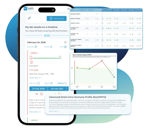
Lab Results Explained and Tracked
What does it mean if your Transferrin result is too high?
Persistent high transferrin most commonly reflects iron deficiency, although pregnancy, estrogen therapy, and oral contraceptives can also increase transferrin production. The liver produces more transferrin to maximize iron capture when iron becomes scarce. This inverse relationship is one of transferrin's most diagnostically important features: transferrin rises as iron falls. A high transferrin result alone does not diagnose iron deficiency anemia; it signals that further evaluation is warranted. The next steps are confirming low serum iron and low ferritin (which would deplete together with developing iron deficiency) and calculating transferrin saturation, which should be low (below 20%) if iron deficiency is the cause. Other causes of elevated transferrin include pregnancy (a physiological rise in the second and third trimesters driven by increased iron demand), oral contraceptive use (estrogen stimulates hepatic transferrin synthesis), and the recovery phase of viral hepatitis. In the absence of these explanations, a persistently elevated transferrin with low ferritin and low serum iron is the classic laboratory picture of iron deficiency and should prompt investigation of its underlying cause — dietary insufficiency, malabsorption (celiac disease, gastrointestinal surgery), or blood loss (gastrointestinal bleeding, heavy menstruation).
Related Health Conditions
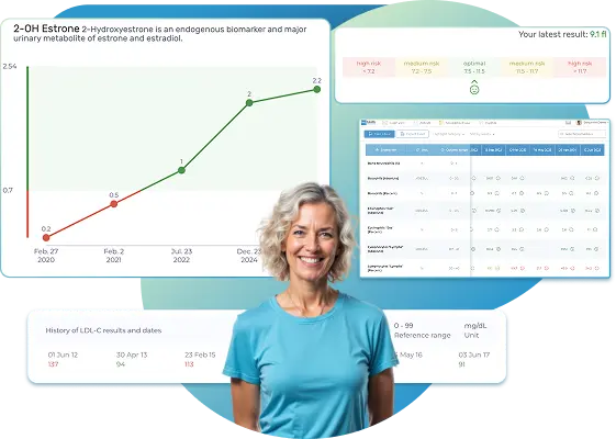
All Your Lab Results.
One Simple Dashboard.
Import, Track, and Share Your Lab Results Easily
Import, Track, and Share Your Lab Results
Import lab results from multiple providers, track changes over time, customize your reference ranges, and get clear explanations for each result. Everything is stored securely, exportable in one organized file, and shareable with your doctor—or anyone you choose.
Cancel or upgrade anytime
What does it mean if your Transferrin result is too low?
Low transferrin (below 200 mg/dL or 2.0 g/L) signals that the liver is producing less of this protein than normal, or that iron-binding sites are so fully occupied that the body is suppressing production. These two mechanisms require opposite clinical responses — one points toward liver, nutrition, or inflammation; the other points toward iron overload. The single most practically useful feature of transferrin in this context is its half-life: at 8–10 days, it responds to changes in nutritional status or liver function roughly twice as fast as albumin (20 days) and recovers faster with treatment. This makes transferrin a sensitive early warning signal for protein malnutrition — when a patient is being nutritionally rehabilitated after critical illness, surgery, or severe dietary restriction, a rising transferrin is often the first measurable laboratory sign of response. The corollary also applies: during acute infection, surgery, or trauma, transferrin falls rapidly as part of the acute-phase response — within 24–48 hours. A low transferrin drawn during an acute hospital admission should always be reinterpreted after recovery, since it may normalize completely once inflammation resolves. Distinguishing these scenarios from iron overload and liver disease requires checking ferritin (elevated in iron overload), CRP (elevated in acute inflammation), albumin (falls in parallel with transferrin in liver disease and malnutrition but not in isolated iron overload), and transferrin saturation (high in iron overload, often normal or low in the other causes).
Related Biomarkers
- Albumin, Serum
- C-Reactive Protein (CRP)
- Ferritin
- Ferritin (female range)
- Hemoglobin
- Hemoglobin (Female range)
- Iron
- Mean Corpuscular Hemoglobin (MCH)
- Mean Corpuscular Volume (MCV)
- RDW-CV (Red Cell Distribution Width) in %
- RDW-SD (Red Cell Distribution Width) in fL
- Soluble Transferrin Receptor
- Total iron-binding capacity (TIBC)
- Transferrin saturation (Iron Saturation)
- Unsaturated Iron Binding Capacity (UIBC)
Article Review & Sources
All our content is backed by peer-reviewed studies, academic research, and trusted medical sources. We're committed to accuracy and transparency — see our editorial policy for details.
Laboratories
Bring All Your Lab Results Together — In One Place
We accept reports from any lab, so you can easily collect and organize all your health information in one secure spot.


Pricing Table
Gather Your Lab History — and Finally Make Sense of It
Finally, Your Lab Results Organized and Clear
Personal plans
$79/ year
Advanced Plan
Access your lab reports, explanations, and tracking tools.
- Import lab results from any provider
- Track all results with visual tools
- Customize your reference ranges
- Export your full lab history anytime
- Share results securely with anyone
- Receive 5 reports entered for you
- Cancel or upgrade anytime
$250/ once
Unlimited Account
Pay once, access everything—no monthly fees, no limits.
- Import lab results from any provider
- Track all results with visual tools
- Customize your reference ranges
- Export your full lab history anytime
- Share results securely with anyone
- Receive 10 reports entered for you
- No subscriptions. No extra fees.
$45/ month
Pro Monthly
Designed for professionals managing their clients' lab reports
- Import lab results from any provider
- Track lab results for multiple clients
- Customize reference ranges per client
- Export lab histories and reports
- Begin with first report entered by us
- Cancel or upgrade anytime
About membership
What's included in a Healthmatters membership
 Import Lab Results from Any Source
 See Your Health Timeline
 Understand What Your Results Mean
 Visualize Your Results
 Data Entry Service for Your Reports
 Securely Share With Anyone You Trust
Let Your Lab Results Tell the Full Story
Once your results are in one place, see the bigger picture — track trends over time, compare data side by side, export your full history, and share securely with anyone you trust.


Bring all your results together to compare, track progress, export your history, and share securely.
What Healthmatters Members Are Saying

I have been using Healthmatters.io since 2021. I travel all over the world and use different doctors and health facilities. This site has allowed me to consolidate all my various test results over 14 years in one place. And every doctor that I show this to has been impressed. Because with any health professional I talk to, I can pull up historical results in seconds. It is invaluable. Even going back to the same doctor, they usually do not have the historical results from their facility in a graph format. That has been very helpful.
Anthony
Unlimited Plan Member since 2021
What fantastic service and great, easy-to-follow layouts! I love your website; it makes it so helpful to see patterns in my health data. It's truly a pleasure to use. I only wish the NHS was as organized and quick as Healthmatters.io. You've set a new standard for health tracking!
Karin
Advanced Plan Member since 2020
As a PRO member and medical practitioner, Healthmatters.io has been an invaluable tool for tracking my clients' data. The layout is intuitive, making it easy to monitor trends and spot patterns over time. The ability to customize reports and charts helps me present information clearly to my clients, improving communication and outcomes. It's streamlined my workflow, saving me time and providing insights at a glance. Highly recommended for any practitioner looking for a comprehensive and user-friendly solution to track patient labs!
Paul
Healthmatters Pro Member since 2024
We implement proven measures to keep your data safe.
At HealthMatters, we're committed to maintaining the security and confidentiality of your personal information. We've put industry-leading security standards in place to help protect against the loss, misuse, or alteration of the information under our control. We use procedural, physical, and electronic security methods designed to prevent unauthorized people from getting access to this information. Our internal code of conduct adds additional privacy protection. All data is backed up multiple times a day and encrypted using SSL certificates. See our Privacy Policy for more details.
