
RBC in Stool: What 0-1, 1-2 and 2-4 per HPF Mean and Whether a Normal Range Exists
Other names: RBC, RBCs, Red Blood Cells, Red Blood Cell, Erythrocytes, RBC Stool, RBC in Stool, RBCs in Stool, Red Blood Cells Stool, Stool RBC, Fecal RBC, RBC Fecalysis, RBC in Fecalysis, Fecalysis RBC, Red Blood Cells Fecalysis, RBC Stool Microscopy, Stool RBC Microscopy, RBC HPF Stool, RBC per HPF, RBC/HPF, Erythrocytes Stool, Erythrocytes Presence Stool, RBC Stool Exam, Stool Exam RBC, Microscopic RBC Stool, GI360 RBC, Doctor's Data RBC Stool
 Reference range:

At a Glance
Red blood cells in stool are looked for under a microscope, and laboratories report what was seen in two different formats. This Doctor's Data panel reports an abundance category: Not Detected, Few, or Moderate. Hospital and clinic laboratories report a number or a range of red cells per high power field instead — 0-1/HPF, 1-2/HPF, 2-4/HPF — where HPF means one view through the microscope at high magnification.
Red cells are not an expected constituent of stool, so blood reaching the sample is a finding to explain. But no single cutoff per high power field applies across laboratories, and none of the authorities behind this page defines one. What a number means, and whether your laboratory flags it, depends on its method and its own reference information.
What the number can and cannot be read as
The two formats are not interchangeable. An abundance category such as Few is a judgment of how readily red cells were found; a figure such as 1-2/HPF is a count of red cells per microscope field. A count is a measurement — but of cells in the fields examined, not of blood lost. Neither format is calibrated against a volume, so neither tells you how much blood there is.
Neither locates the bleeding. Intact red cells break down on a long passage through the gut, a reason their presence tends to indicate blood entering lower down — but a tendency is not a localization.
The LOINC concept these results are filed under carries an ordinal scale, which is what this panel's Not Detected / Few / Moderate reporting matches. LOINC is explicit that an ordinal scale says nothing about whether a laboratory used an internal threshold to reach the category, so the coding is not evidence about cutoffs either way — and it describes that concept's reporting scale, not the procedures of every laboratory offering stool microscopy.
How to read your result
Read the result next to the rest of the report and next to how you feel, not on its own.
Not Detected, or 0/HPF, says no intact red cells were seen in the fields examined. It does not say there is no blood in your stool, and it is not a negative cancer screen.
A low number such as 0-1/HPF or 0-2/HPF is a low count per high power field. How that figure or range is generated, and whether it is flagged, is set by the reporting laboratory — check the reference information on your own report.
Few, or 1-2/HPF and 2-4/HPF, says intact red cells were seen. Moderate, or a higher count, says they were seen more readily. This result alone does not determine urgency and does not establish a diagnosis.
When to seek care regardless of the category
Urgency is decided by symptoms, not by this line. Seek help right away for signs of acute or severe gastrointestinal bleeding — black tarry stools, large amounts of visible blood, faintness or dizziness. The signs of shock are an emergency: confusion or loss of consciousness, a fast heart rate, pale skin, cold hands and feet, sweating. That guidance is the same whether this line reads Not Detected, Few or Moderate.
If you remember only one thing
A low number is not a graded mild abnormality, and a result of none is not an all-clear. This observation says whether intact red cells were seen and roughly how many — not how much blood there is, and not where it came from. A negative result does not exclude bleeding, including bleeding the occult blood test beside it might detect.
RBC microscopy and occult blood: what each one detects
On this panel, Red Blood Cells and Occult Blood are separate analytes and they detect different things. Microscopy looks for intact cells. Occult blood assays look for blood components — heme or globin — which persist after the cells have broken down. That is why blood can register on one and not the other.
It is tempting to turn that into a map of the gut, and worth resisting. Doctor's Data's guide does say an elevated red cell result "indicates active bleeding lower in the intestinal tract," and that positive occult blood "in many cases" comes from "the upper small intestine or higher." But the same paragraph goes on to name colon cancer, ulcerative colitis, diverticulitis, and ulceration of the esophagus, stomach or duodenum. Colonic causes appear on both markers; their coverage overlaps. Occult blood assays also differ by method in what they detect and where they are most sensitive, and the guide does not state which method this panel uses, so nothing method-specific should be read into either result.
The practical rule is short. Read both, because either can register blood the other misses. Neither localizes bleeding, and a negative result on either — or both — does not exclude it when symptoms or your own observations warrant assessment.
Is this a colon cancer screening test?
No, and it should not be used as one.
The US Preventive Services Task Force recommends colorectal cancer screening for asymptomatic average-risk adults aged 50 to 75 (grade A) and 45 to 49 (grade B), and names the stool-based options: annual high-sensitivity guaiac fecal occult blood testing, annual fecal immunochemical testing, or stool DNA-FIT every one to three years. Microscopy for red cells is not one of them.
Two things follow. A Not Detected result here is not a negative cancer screen and does not substitute for one. And a Few or Moderate result is not a cancer finding — it is a finding that blood was seen, which has many causes.
Two people, same result
Two people both get Few.
The first is 26, has had visible blood on the paper for a month, and a painful fissure. The blood is coming from a few centimeters inside the outlet, and the microscopy is telling her what she already knows.
The second is 71, has had looser stools for three months, has lost weight, and is anemic. The same Few sits in a different picture. In a population-based series of patients investigated for rectal bleeding, diverticular bleeding accounted for 33.1% of cases, hemorrhoidal disease 20%, colorectal polyps 13.1%, colorectal malignancy 8.2%, ischemic colitis 6.6% and angiodysplasia 6% — and the finding carries more weight when bleeding comes with older age, weight loss, altered bowel habits or severe anemia.
The marker is identical. What it is worth is not.
What this result cannot tell you
Where the blood came from, or how much there is. Neither is answerable from this line.
A cause. The 2017 Infectious Diseases Society of America guideline is explicit about the paired microscopy marker: fecal leukocyte examination and stool lactoferrin detection should not be used to establish the cause of acute infectious diarrhea (strong recommendation, moderate evidence), and it found insufficient data to recommend fecal calprotectin there either. Red cell microscopy is not a more powerful instrument than the leukocyte examination beside it.
And it rules nothing out — not cancer, not bleeding.
What makes red cells appear in stool
Doctor's Data's comment for an elevated result names four groups: enteroinvasive bacterial or parasitic infection, an active inflammatory bowel condition such as ulcerative colitis, hemorrhoids — which the guide calls "very common" — and colorectal cancer or anal fistulas. NIDDK adds diverticular disease, anal fissures, polyps, and colitis among causes of lower gastrointestinal bleeding.
On the infection route the laboratory points to its own PCR and culture, naming Shigella, Salmonella, Campylobacter and Yersinia, which cause mucosal ulceration. The IDSA guideline's list of pathogens producing visible blood in stool is wider: Shiga toxin-producing E. coli, Shigella, Salmonella, Campylobacter, Entamoeba histolytica, non-cholera Vibrio species, Yersinia, Balantidium coli and Plesiomonas.
For the inflammatory route, the laboratory's instruction is to check fecal calprotectin and lactoferrin, both on this same panel. Raised results there support intestinal inflammation without identifying its cause: Mayo Clinic Laboratories states that elevated fecal calprotectin is not diagnostic for inflammatory bowel disease, that normal concentrations do not exclude it, and names celiac disease, colorectal cancer and gastrointestinal infections among other causes.
One observation on this panel is a red cell finding of a different kind. The trichrome parasitology looks for amebic trophozoites, and red cells inside a trophozoite's cytoplasm are classically associated with Entamoeba histolytica — but CDC states this may rarely occur with E. dispar too, so it "is not a reliable criterion by which to distinguish these species," that it is rarely seen on stained smears, and that molecular methods are what it recommends for telling these species apart. Free red cells in the smear and red cells inside a parasite are separate observations, and the second needs species-specific testing behind it.
Common interpretation mistakes
Reading the result as a quantity of blood. Neither reporting format is calibrated against volume.
Reading Not Detected as a reason to stop. It says no intact red cells were seen, not that there is no blood and not that nothing is wrong.
Reading a low number as confirmed-normal, or confirmed-abnormal. Whether your laboratory flags it is set by its method and reference information, printed on your report.
Letting the result decide urgency in either direction. Symptoms decide that.
Using this marker in place of a colorectal cancer screening test, or reading a negative result as a negative screen.
Treating everything that puts blood in the sample as the same kind of event. Menstrual blood contaminates the specimen. Bleeding hemorrhoids or an anal fissure are genuine sources near the outlet — real findings, not artifacts to discount. Either way, mention the collection circumstances to whoever ordered the test, and follow that laboratory's instructions.
Reading an adult interval onto a child's result. Doctor's Data states for this panel that "the majority of reference intervals are established from adult populations. Results may differ in pediatric populations and care should be taken when interpreting these values."
Questions your doctor may ask
Have you seen blood yourself, and was it bright red, dark, or mixed through the stool? Was there pain passing it? Any change in the shape, frequency or looseness of your stools, and for how long? Any unintended weight loss, faintness or dizziness? Any fever, recent travel, or anyone else ill around you? Were you menstruating, or were your hemorrhoids bleeding, when you collected the sample? Are you taking aspirin, an anti-inflammatory, or a blood thinner? Any family history of colorectal cancer or inflammatory bowel disease, and have you had a colonoscopy?
Read together with
Occult Blood is the first to read, because it detects blood components rather than cells and can register blood this marker misses — the reverse is also true, and neither locates the source. White Blood Cells indicates inflammation rather than bleeding. Calprotectin and Lactoferrin are the quantitative inflammation markers, and the two the laboratory's guide tells you to check when an inflammatory bowel condition is in question.
The rest of the microscopy section sits alongside: Mucus, Muscle Fibers, Vegetable Fibers, microscopic yeast, and Charcot-Leyden Crystals, which form from eosinophil breakdown and can appear in parasitic disease.
The GI Pathogens PCR panel on the same report is the instrument that can actually name an infectious cause, which the microscopy cannot. The parasitology preparations sit beside it.
Away from the stool panel, a complete blood count can assess anemia, measuring red blood cell count, hemoglobin and hematocrit among other things. Ferritin is a separate test — not part of a complete blood count — and one of the iron studies for iron deficiency; inflammation raises it, so it is read in context. Neither identifies a bleeding source, and normal results do not exclude bleeding.
Clinical pearls
An abundance category is not a low-resolution count. Converting this panel's Few into an implied number, or a count per field into an implied volume of blood, adds precision the observation never had.
A tendency is not a localization. Intact red cells surviving to the sample is a reason to think of sources lower down; it is not an answer about where the blood came from, and the occult blood marker does not answer it either.
Red cells and white cells together, in acute diarrhea, is a reason to look at the pathogen testing on the same report — not a reason to conclude the infection has been identified.
Clinical Takeaway
Red cells are not an expected constituent of stool, so their presence is a finding to explain. But no universal cutoff per high power field appears in the sources behind this page, and whether your laboratory flags a low number is set by its own method and reference information, printed on your report. Whichever format you have, the result does not measure blood loss, locate bleeding, establish a cause, or serve as a cancer screen, and a negative result does not exclude bleeding. Read it beside Occult Blood, White Blood Cells, Calprotectin and Lactoferrin, and let symptoms decide how quickly to act.
In one sentence
Stool RBC microscopy reports whether intact red cells were seen and roughly how abundant they were; it does not measure blood loss or identify its source.
Bottom line
Take the report to a clinician if red cells were seen, if you have seen blood yourself, or if your stools have changed — and seek help right away, whatever this line says, for black tarry stools, heavy bleeding, faintness, or signs of shock.
FAQ about RBC
-
What is the normal range for RBC in stool?
Red cells are not an expected constituent of stool, so blood reaching the sample is a finding to explain rather than a normal variant. Beyond that, there is no single cutoff for red cells per high power field that applies across laboratories, and none of the authorities behind this page defines one. What a given number means, and whether it is flagged, is set by the reporting laboratory's method and its own reference information — which is printed on your report and is the thing to check. The LOINC concept these results are filed under, Erythrocytes [Presence] in Stool, carries an ordinal scale, meaning results against that concept are reported as an ordered category. LOINC is explicit that this says nothing either way about whether a laboratory used an internal threshold to reach the category, so the coding is not evidence about cutoffs in either direction. That code matches this Doctor's Data panel's categorical reporting; it does not establish how another laboratory counts or reports cells per high power field. -
What does RBC 0-1/HPF in stool mean?
It is a low reported number of red blood cells per high power field. HPF stands for high power field — one circular view through the microscope at high magnification. How a laboratory arrives at a range rather than a single figure depends on its own reporting procedure, and nothing in the sources behind this page defines that procedure, so the endpoints should not be read as a minimum and maximum across fields. The same applies to 0-2/HPF. What this number means for you, and whether your laboratory flags it, depends on that laboratory's method and reference information, so the reference range printed on your own report is the thing to look at rather than a figure from elsewhere. Two limits hold whatever the number: it is not calibrated against a volume, so it does not measure blood loss, and it does not tell you where the blood came from. What matters alongside it is whether you have seen blood yourself, whether your stools have changed, and what the other markers on the report show — particularly Occult Blood, which detects blood components the microscopy cannot see. -
What does RBC 1-2/HPF or 2-4/HPF in stool mean?
It means intact red blood cells were seen in the fields examined. The two figures are the laboratory's reported range of red cells per high power field; how that range is generated depends on its reporting procedure. Red cells are not an expected constituent of stool, so this says blood reached the sample. It does not say how much, and it does not say where it came from. Intact red cells break down during a long passage through the gut, which is a reason their presence tends to indicate blood entering lower down — a fissure or hemorrhoid near the outlet, a diverticulum, an inflamed or ulcerated patch of lining, a polyp, or an invasive infection — but a tendency is not a localization. Two collection circumstances are worth mentioning to whoever ordered the test, and they are different things: menstrual blood contaminates the specimen, while bleeding hemorrhoids or an anal fissure are genuine sources of blood near the outlet. This result alone does not determine urgency; symptoms do. -
What does RBC mean on a fecalysis result?
Fecalysis is a routine stool examination, and the RBC line is the microscopic search for intact red blood cells in the sample. It is the same observation described elsewhere as stool RBC microscopy or red blood cells in stool. Fecalysis reports tend to give it as a count per high power field, such as 0-1/HPF or 1-2/HPF, while specialty stool panels such as this one use ordered words instead — Not Detected, Few, Moderate. Both answer the same question: whether red cells were seen, and roughly how many. Neither is a measurement of blood loss, and what a given figure means is set by the reporting laboratory's own method and reference information. The WBC line beside it counts white blood cells and indicates inflammation rather than bleeding, and the two are read together: red cells and white cells appearing together in someone with acute diarrhea points toward an invasive infection and toward the pathogen testing that can actually identify one. -
Is RBC in stool the same as a positive occult blood test?
No. They are separate tests, reported separately, and on this panel they are separate analytes that detect different things. Microscopy looks for intact cells. Occult blood assays look for blood components — heme or globin — which persist after the cells have broken down. That is why blood can register on one and not the other. It is tempting to turn this into a map of the gut, and worth resisting: Doctor's Data's guide does say an elevated red cell result indicates bleeding lower in the intestinal tract and that positive occult blood in many cases comes from the upper small intestine or higher, but the same occult blood paragraph goes on to name colon cancer, ulcerative colitis and diverticulitis, which are colonic. The coverage overlaps. Read both, because either can register blood the other misses, and treat neither as locating the bleeding or as excluding it. -
Does RBC in stool mean I have colon cancer?
No. Red cells seen in stool means blood reached the sample, and blood in stool has many causes. In a population-based series of patients investigated for rectal bleeding, diverticular bleeding accounted for 33.1% of cases, hemorrhoidal disease 20%, colorectal polyps 13.1%, colorectal malignancy 8.2%, ischemic colitis 6.6% and angiodysplasia 6%. It also works the other way, and this matters more: a result of none is not a negative cancer screen. Stool microscopy for red cells is not a colorectal cancer screening test. The US Preventive Services Task Force recommends screening for asymptomatic average-risk adults aged 50 to 75 and 45 to 49, and the stool-based options it names are annual fecal immunochemical testing, annual high-sensitivity guaiac fecal occult blood testing, or stool DNA-FIT every one to three years. If you are due for screening, this line does not count toward it. -
Can RBC in stool come from something other than bleeding in my gut?
Yes, though the possibilities are not all the same kind of thing. Menstrual blood can contaminate the specimen — that is blood in the sample rather than blood from your gut. Bleeding hemorrhoids and anal fissures are different: they are genuine sources of blood entering near the outlet, and NIDDK lists both among the causes of lower gastrointestinal bleeding. So a result explained by hemorrhoids is still explained by real bleeding, just from a place that is usually benign. The University of Iowa laboratory handbook instructs that its guaiac occult blood test should not be performed with actively bleeding hemorrhoids or during menstruation; that instruction is written for that test rather than for red cell microscopy, so the thing to follow is the collection guidance from the laboratory that ran yours. Either way, the timing and circumstances of the collection belong in the conversation with whoever ordered the test, and a repeat sample taken at a different time is sometimes the clearest next step. -
What should I check next on the same report?
Four things sit alongside it. Occult Blood detects blood components rather than cells, so it can register blood the microscopy misses — the reverse is also true, and neither locates the source. White Blood Cells indicates inflammation rather than bleeding. Calprotectin and Lactoferrin are the quantitative inflammation markers: a raised result on either alongside red cells supports intestinal inflammation, but does not distinguish inflammatory bowel disease from infection or other causes — Mayo Clinic Laboratories states that elevated calprotectin is not diagnostic for IBD, that a normal result does not exclude it, and that celiac disease, colorectal cancer and gastrointestinal infections can also raise it. The laboratory's own comment on this marker still directs you to those two when an inflammatory bowel condition is being considered. The GI Pathogens PCR panel is what can name an infectious cause; the microscopy cannot. Away from the stool panel, a complete blood count can assess anemia, and ferritin — a separate test, not part of a complete blood count — is one of the iron studies for iron deficiency, read in light of the fact that inflammation raises it. -
Why does my report say Few or Moderate instead of a number?
Because this Doctor's Data panel reports approximate abundance using ordered categories, and the wording matches that. The LOINC concept these results are filed under carries an ordinal scale, which is the same shape, and Doctor's Data uses that style across its microscopy section; for microscopic yeast its guide describes small quantities as reported as rare and higher amounts as moderate to many. A laboratory that prints 1-2/HPF has made a different choice: that is a count of cells per microscope field, which is a measurement, though of cells in the fields examined rather than of blood lost. Neither format converts into the other, and neither converts into a volume of blood. The step from Few to Moderate is a step in how readily red cells were seen, not a calibrated step in blood loss. Read either format the same way: were red cells seen, roughly how many, what does the reference information on your own report say about it, and what do the rest of the report and your own symptoms say alongside it.
Lab Results Explained and Tracked
Related Health Conditions

All Your Lab Results.
One Simple Dashboard.
Import, Track, and Share Your Lab Results Easily
Import, Track, and Share Your Lab Results
Import lab results from multiple providers, track changes over time, customize your reference ranges, and get clear explanations for each result. Everything is stored securely, exportable in one organized file, and shareable with your doctor—or anyone you choose.
Cancel or upgrade anytime
Related Biomarkers
Article Review & Sources
All our content is backed by peer-reviewed studies, academic research, and trusted medical sources. We're committed to accuracy and transparency — see our editorial policy for details.
Laboratories
Bring All Your Lab Results Together — In One Place
We accept reports from any lab, so you can easily collect and organize all your health information in one secure spot.











Pricing Table
Gather Your Lab History — and Finally Make Sense of It
Finally, Your Lab Results Organized and Clear

Personal plans

$79/ year
Advanced Plan
Access your lab reports, explanations, and tracking tools.
- Import lab results from any provider
- Track all results with visual tools
- Customize your reference ranges
- Export your full lab history anytime
- Share results securely with anyone
- Receive 5 reports entered for you
- Cancel or upgrade anytime
$250/ once
Unlimited Account
Pay once, access everything—no monthly fees, no limits.
- Import lab results from any provider
- Track all results with visual tools
- Customize your reference ranges
- Export your full lab history anytime
- Share results securely with anyone
- Receive 10 reports entered for you
- No subscriptions. No extra fees.
$45/ month
Pro Monthly
Designed for professionals managing their clients' lab reports
- Import lab results from any provider
- Track lab results for multiple clients
- Customize reference ranges per client
- Export lab histories and reports
- Begin with first report entered by us
- Cancel or upgrade anytime
About membership
What's included in a Healthmatters membership
 Import Lab Results from Any Source
 See Your Health Timeline
 Understand What Your Results Mean

 Visualize Your Results

 Data Entry Service for Your Reports
 Securely Share With Anyone You Trust
Let Your Lab Results Tell the Full Story
Once your results are in one place, see the bigger picture — track trends over time, compare data side by side, export your full history, and share securely with anyone you trust.




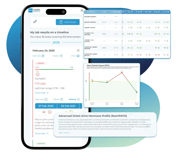
Bring all your results together to compare, track progress, export your history, and share securely.
What Healthmatters Members Are Saying

I have been using Healthmatters.io since 2021. I travel all over the world and use different doctors and health facilities. This site has allowed me to consolidate all my various test results over 14 years in one place. And every doctor that I show this to has been impressed. Because with any health professional I talk to, I can pull up historical results in seconds. It is invaluable. Even going back to the same doctor, they usually do not have the historical results from their facility in a graph format. That has been very helpful.
Anthony
Unlimited Plan Member since 2021
What fantastic service and great, easy-to-follow layouts! I love your website; it makes it so helpful to see patterns in my health data. It's truly a pleasure to use. I only wish the NHS was as organized and quick as Healthmatters.io. You've set a new standard for health tracking!
Karin
Advanced Plan Member since 2020
As a PRO member and medical practitioner, Healthmatters.io has been an invaluable tool for tracking my clients' data. The layout is intuitive, making it easy to monitor trends and spot patterns over time. The ability to customize reports and charts helps me present information clearly to my clients, improving communication and outcomes. It's streamlined my workflow, saving me time and providing insights at a glance. Highly recommended for any practitioner looking for a comprehensive and user-friendly solution to track patient labs!
Paul
Healthmatters Pro Member since 2024

We implement proven measures to keep your data safe.
At HealthMatters, we're committed to maintaining the security and confidentiality of your personal information. We've put industry-leading security standards in place to help protect against the loss, misuse, or alteration of the information under our control. We use procedural, physical, and electronic security methods designed to prevent unauthorized people from getting access to this information. Our internal code of conduct adds additional privacy protection. All data is backed up multiple times a day and encrypted using SSL certificates. See our Privacy Policy for more details.