
Anti-Ma (IgG + IgA)

Optimal result: 0 – 10 Relative Abundance

The Anti-Ma (IgG + IgA) biomarker is a diagnostic marker used to detect autoantibodies against Ma antigens, which are proteins found in the brain, particularly in the Purkinje cells of the cerebellum. These antibodies are typically associated with autoimmune responses that target the central nervous system (CNS). The Anti-Ma test, often part of a broader neurological autoantibody panel like Vibrant America’s Neural Zoomer Plus, is crucial for identifying potential paraneoplastic syndromes and other neurological autoimmune disorders.

-----------

If Anti-Ma (IgG + IgA) is mildly elevated, it may suggest an autoimmune response targeting Ma antigens, which are primarily found in neurons, particularly in the Purkinje cells of the cerebellum. These antibodies are often associated with paraneoplastic syndromes (neurological conditions triggered by an underlying malignancy) and other autoimmune neurological disorders.

Possible Implications of Mild Elevation:

  1. Paraneoplastic Cerebellar Degeneration (PCD):

    • Anti-Ma (IgG + IgA) antibodies are commonly found in paraneoplastic cerebellar degeneration, a condition where the immune system attacks the cerebellum in response to cancer, particularly ovarian, breast, and small-cell lung cancer.

    • Mild elevation may still be indicative of a paraneoplastic syndrome, even if the patient does not have overt symptoms or a diagnosed malignancy. Further cancer screening (e.g., imaging, PET scans) may be warranted to rule out an underlying tumor.

  2. Autoimmune Neurological Disorders:

    • In some cases, Anti-Ma antibodies can be present in autoimmune cerebellar ataxia, a condition where the immune system attacks the cerebellum without the presence of cancer. Mildly elevated levels may indicate an autoimmune process affecting the central nervous system (CNS).

  3. Early or Subclinical Autoimmune Response:

    • A mildly elevated Anti-Ma (IgG + IgA) level could represent an early-stage autoimmune reaction that has not yet developed into a more severe condition. This could be indicative of a developing autoimmune neurological disorder, which may progress if not addressed.

    • Monitoring levels over time, along with the appearance of symptoms, can help track the progression of the autoimmune response.

  4. Non-specific or False Positive:

    • A mildly elevated Anti-Ma (IgG + IgA) could also be due to non-specific immune activation or cross-reactivity with other antigens. In such cases, further testing, such as CSF analysis or additional autoimmune panels, may help clarify the cause.

    • It’s important to correlate the antibody results with the patient's clinical history and symptoms.

Next Steps in Evaluation:

  • Clinical Correlation: The elevation should be interpreted in light of any symptoms the patient may be experiencing, such as motor coordination difficulties (e.g., ataxia), dysarthria (difficulty speaking), or nystagmus (involuntary eye movements). These symptoms may suggest a neurological condition, possibly linked to paraneoplastic syndrome or an autoimmune disorder.

  • Cancer Screening:
    If Anti-Ma (IgG + IgA) levels are mildly elevated, it’s crucial to rule out an underlying malignancy. This could involve:

    • Imaging (e.g., CT scans, MRI, PET scans) to detect tumors, especially ovarian, breast, or lung cancers.

    • Tumor marker tests to identify cancer-related autoimmunity.

  • Additional Autoimmune Testing:
    Further tests, such as CSF analysis (looking for intrathecal antibody production), Anti-Yo, Anti-Hu, or Anti-Ri antibodies, may help confirm a diagnosis of paraneoplastic cerebellar degeneration or another neurological autoimmune condition.

Conclusion:

A mildly elevated Anti-Ma (IgG + IgA) level suggests that there may be an autoimmune response affecting the nervous system, possibly related to paraneoplastic cerebellar degeneration or another autoimmune condition. The next steps typically involve further testing, such as cancer screening and additional autoimmune panels, to clarify the cause and guide appropriate treatment. Monitoring symptoms and antibody levels over time is important for detecting any progression of the condition.

The Anti-Ma (IgG + IgA) biomarker is a diagnostic marker used to detect autoantibodies against Ma antigens, which are proteins found in the brain, particularly in the Purkinje cells of the cerebellum. These antibodies are typically associated with autoimmune responses that target the central nervous system (CNS). The Anti-Ma test, often part of a broader neurological autoantibody panel like Vibrant America’s Neural Zoomer Plus, is crucial for identifying potential paraneoplastic syndromes and other neurological autoimmune disorders.

What It Measures

The Anti-Ma (IgG + IgA) test detects IgG and IgA antibodies directed against Ma antigens, which are involved in neuronal function. These antibodies can target intracellular or membrane-associated proteins in neurons, particularly those related to the cerebellum and brainstem, and are considered a hallmark of certain paraneoplastic neurological disorders.

Clinical Relevance

Anti-Ma antibodies are strongly associated with paraneoplastic cerebellar degeneration (PCD) and other autoimmune neurological conditions. These conditions occur when the immune system mistakenly attacks the nervous system, often in response to an underlying malignancy. Anti-Ma (IgG + IgA) testing is particularly relevant for diagnosing the following:

  • Paraneoplastic Cerebellar Degeneration (PCD):

    • This condition is often associated with cancers, particularly ovarian, breast, and small-cell lung cancer. Anti-Ma antibodies are found in a significant percentage of patients with PCD and are commonly linked to cerebellar ataxia, which affects motor coordination and balance.

    • Patients may present with subacute cerebellar ataxia, dysarthria (speech difficulty), and nystagmus (involuntary eye movements).

  • Paraneoplastic Syndromes:

    • These are a group of disorders that occur when the immune system targets healthy tissue, often triggered by the presence of cancer. Anti-Ma antibodies can be present in paraneoplastic neurological syndromes, especially those affecting the cerebellum and brainstem.

  • Neurodegenerative Diseases:

    • Some research suggests that Anti-Ma antibodies may also be present in neurodegenerative diseases, though these cases are less common. The role of Anti-Ma in such conditions is still under investigation.

Interpretation of Results

  • Positive Result (IgG/IgA):

    • A positive test for Anti-Ma antibodies suggests an autoimmune attack on the nervous system, particularly in the cerebellum. This is commonly seen in paraneoplastic cerebellar degeneration or other autoimmune conditions affecting the CNS.

    • Further testing, including cancer screening (e.g., CT scans, PET scans), is recommended to rule out an underlying malignancy. If a malignancy is found, treating the cancer often leads to stabilization or improvement of neurological symptoms.

  • Negative Result:

    • A negative result typically indicates that Anti-Ma antibodies are not present, which reduces the likelihood of paraneoplastic cerebellar degeneration but does not exclude other autoimmune or neurodegenerative conditions.

    • However, the absence of Anti-Ma antibodies does not rule out the presence of paraneoplastic syndromes, as other autoantibodies may be involved in different presentations of these disorders.

Testing Considerations

  • Panel Context: The Anti-Ma (IgG + IgA) test is typically part of a broader neuroautoantibody panel designed to evaluate various antibodies that may contribute to neurological symptoms such as balance issues, muscle weakness, and cognitive dysfunction. It is often used in conjunction with other biomarkers like Anti-Yo, Anti-Hu, and Anti-Ri, which are also associated with paraneoplastic syndromes.

  • Complementary Tests:

    • Cancer Screening: A positive result for Anti-Ma antibodies may prompt further testing to identify any underlying cancer, especially ovarian, breast, or lung cancer, which are most commonly associated with paraneoplastic cerebellar degeneration.

    • CSF Analysis: For patients with neurological symptoms, cerebrospinal fluid (CSF) analysis can provide additional insight into the presence of autoimmune activity in the CNS.

  • Limitations:

    • The presence of Anti-Ma antibodies is not always indicative of paraneoplastic cerebellar degeneration or malignancy. In some cases, these antibodies may be present in non-cancerous autoimmune conditions or neurodegenerative diseases.

    • False positives can also occur in patients with other autoimmune conditions, so clinical correlation with symptoms is essential for accurate diagnosis.

Treatment Implications

  • Immunotherapy: For autoimmune conditions, particularly paraneoplastic cerebellar degeneration, immunosuppressive therapies such as IVIg, steroids, or plasmapheresis may be used to reduce the immune response and alleviate symptoms.

  • Cancer Treatment: If a malignancy is identified, tumor resection or other cancer treatments may help improve neurological symptoms associated with paraneoplastic syndromes.

Conclusion

Anti-Ma (IgG + IgA) testing is an important tool for diagnosing autoimmune neurological disorders, particularly paraneoplastic cerebellar degeneration. A positive result suggests an autoimmune response against the cerebellum, often triggered by an underlying malignancy. Early detection and treatment are crucial for managing both the neurological symptoms and any associated cancer, making this test a key part of comprehensive neurological care.

Anti-Ma (IgG + IgA)
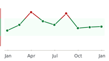
Healthmatters dashboard showing biomarker trend graph

Every lab result you’ve ever had, charted and explained.

Healthmatters is a tracker for your lab results. Upload a report from any lab, any country — every marker charted and explained.

Start with my report →
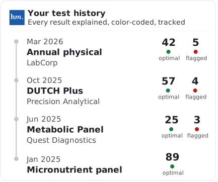
Beyond Anti-Ma (IgG + IgA) — 10,000+ markers explained
One Anti-Ma (IgG + IgA) result tells you where you are. Your history tells you where you’re heading. Upload your past lab reports and see this marker — and every marker on them —every marker charted against your own lab’s ranges.
Start with this report →
Healthmatters timeline preview showing biomarker trends over time
Clinical Sources & References

All our content is backed by peer-reviewed studies, academic research, and trusted medical sources. We’re committed to accuracy and transparency — see our editorial policy for details.

Article Review Log
Written by Healthmatters Editorial Team
Healthmatters Lifetime Plan

Pay $250. Track your health forever

One payment. No renewals. Yours for life.

10,000+ biomarkers explained · trends over time

See other plans →
Secure checkout via Stripe · GDPR & HIPAA
We accept results from any lab
LabCorp Quest Diagnostics Genova Diagnostics DUTCH Test Doctor's Data Vibrant America ZRT Laboratory Cyrex Labs + any other lab

What members are saying

★★★★★ 10,000+ happy members
★★★★★

"Watching my numbers move over the years — instead of forgetting last time's results — completely changed how I manage my health."

Daniel
Member since 2022
★★★★★

"Living across several countries made keeping my lab results organized a real challenge. Healthmatters brought my results from the USA, Spain, and Germany together — 20 years of history, with long-term trends instead of scattered reports."

Ben
Member since 2021 · tracks across countries
★★★★★

"Standard lab ranges are too wide for how I practice — 'normal' isn't optimal. I set my own bands, so a client sees yellow before a marker goes officially out of range. That's the conversation I want to be having."

Dr. Alexandra
Functional medicine · 6 yrs on Pro