
Appearance

Sample type: Synovial fluid
Clear
Normal synovial fluid is clear. Cloudiness or turbidity may suggest the presence of cells, crystals, or other substances.
Hazy
Cloudiness or turbidity may suggest the presence of cells, crystals, or other substances.
Cloudy
Cloudiness or turbidity may suggest the presence of cells, crystals, or other substances. A cloudy appearance in a synovial fluid analysis can signify several underlying conditions or issues within the joint. Here's what it may indicate: Infection -- One of the primary reasons for cloudy synovial fluid is the presence of infection within the joint. Cloudiness often results from an increased white blood cell count (a sign of the body's immune response to infection) and the presence of infectious agents like bacteria. This condition is known as septic arthritis. Inflammation -- Joint inflammation, which can occur due to various causes such as autoimmune disorders like rheumatoid arthritis, gout, or injury, can lead to cloudy synovial fluid. The inflammatory response may cause increased cellular debris in the fluid. Crystals -- Certain crystal-induced arthritis conditions, such as gout or pseudogout, can cause cloudiness in synovial fluid. The crystals themselves can cloud the fluid and trigger an inflammatory response. Hemarthrosis -- In cases of joint bleeding (hemarthrosis), the presence of blood in the synovial fluid can make it appear cloudy or turbid. Trauma -- Trauma or injury to the joint can also result in cloudy synovial fluid due to the release of inflammatory molecules and increased white blood cells as part of the healing process. In summary, a cloudy appearance in synovial fluid typically suggests the presence of inflammation, infection, crystals, or bleeding within the joint. It is a critical clue for healthcare professionals to further investigate the underlying cause of joint symptoms and initiate appropriate treatment.

A synovial fluid analysis involves the examination of the appearance and various physical characteristics of synovial fluid to help diagnose joint-related conditions. Interpreting the appearance in a synovial fluid analysis involves assessing several key aspects:

  1. Color: The color of synovial fluid can provide valuable information. Normal synovial fluid is typically straw-colored or transparent. Cloudy or opaque fluid may indicate inflammation or infection.

  2. Clarity: Normal synovial fluid is clear. Cloudiness or turbidity may suggest the presence of cells, crystals, or other substances.

  3. Viscosity: The viscosity or thickness of the fluid can vary. It may be more viscous in certain joint conditions. Increased viscosity may indicate inflammation.

  4. Volume: Assessing the volume of synovial fluid obtained during aspiration is important. An increased volume may be a sign of joint inflammation, such as in arthritis.

  5. Presence of Debris: The presence of debris or particulate matter in the fluid can be indicative of joint damage or inflammation.

  6. Blood Content: The presence of blood in the synovial fluid, indicated by a pink or red appearance, may suggest bleeding within the joint, trauma, or other conditions.

  7. Lubrication: Synovial fluid's primary function is to lubricate joints. A decrease in lubrication properties may indicate joint problems.

Interpreting the appearance of synovial fluid is just one aspect of a comprehensive synovial fluid analysis. It is usually combined with other laboratory tests and clinical evaluation to determine the underlying cause of joint symptoms, such as arthritis, infection, or crystal-induced arthritis.

Appearance
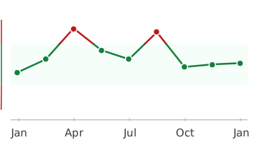
Healthmatters dashboard showing biomarker trend graph

Every lab result you’ve ever had, charted and explained.

Healthmatters is a tracker for your lab results. Upload a report from any lab, any country — every marker charted and explained.

Start with my report →
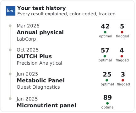
Beyond Appearance — 10,000+ markers explained
One Appearance result tells you where you are. Your history tells you where you’re heading. Upload your past lab reports and see this marker — and every marker on them —every marker charted against your own lab’s ranges.
Start with this report →
Healthmatters timeline preview showing biomarker trends over time
Clinical Sources & References

All our content is backed by peer-reviewed studies, academic research, and trusted medical sources. We’re committed to accuracy and transparency — see our editorial policy for details.

Article Review Log
Written by Healthmatters Editorial Team
Healthmatters Lifetime Plan

Pay $250. Track your health forever

One payment. No renewals. Yours for life.

10,000+ biomarkers explained · trends over time

See other plans →
Secure checkout via Stripe · GDPR & HIPAA
We accept results from any lab
LabCorp Quest Diagnostics Genova Diagnostics DUTCH Test Doctor's Data Vibrant America ZRT Laboratory Cyrex Labs + any other lab

What members are saying

★★★★★ 10,000+ happy members
★★★★★

"Watching my numbers move over the years — instead of forgetting last time's results — completely changed how I manage my health."

Daniel
Member since 2022
★★★★★

"Living across several countries made keeping my lab results organized a real challenge. Healthmatters brought my results from the USA, Spain, and Germany together — 20 years of history, with long-term trends instead of scattered reports."

Ben
Member since 2021 · tracks across countries
★★★★★

"Standard lab ranges are too wide for how I practice — 'normal' isn't optimal. I set my own bands, so a client sees yellow before a marker goes officially out of range. That's the conversation I want to be having."

Dr. Alexandra
Functional medicine · 6 yrs on Pro